ONLINE INQUIRY
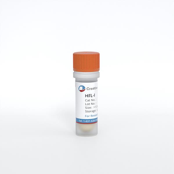
HFL-I
Cat.No.: CSC-C9189W
Species: Human
Source: Lung
Morphology: fibroblast-like
Cell Type: Fibroblast
- Specification
- Background
- Scientific Data
- Q & A
- Customer Review
HFL-I, or HFL-1, is a normal human fibroblast cell line derived from human foetal lung tissue. They are a main component of soft connective tissue, and they derive from mesenchymal cells during embryogenesis. Like other fibroblasts, HFL-I cells exhibit an elongated, spindle-shaped morphology, possess abundant cytoplasm, and have prominent fibrous protrusions.
Embryonic lung fibroblasts contribute to embryonic development, repair and disease. For instance, in development, these cells produce a host of growth factors and cytokines, including transforming growth factor-β (TGF-β), fibroblast growth factor (FGF) and epidermal growth factor (EGF), which regulate alveolar development, differentiation and maturation. They also create and maintain lung tissue's ECM, which is composed of collagen and elastin, both of which not only support the structure of the lungs, but also actively contribute to the expansion and contraction of alveoli. To repair tissue damage, embryonic lung fibroblasts play an important role in correcting cellular decay, necrosis and defects of various severity. In lung diseases such as pulmonary fibrosis and lung cancer, these cells can turn into particular cell types, and help heal and regenerate lung tissue. In disease research, HFL-I is often used to explore mechanisms of pulmonary fibrosis, asthma and chronic obstructive pulmonary disease (COPD). For new therapeutic interventions, a closer examination of the biology and regulation of embryonic lung fibroblasts will be necessary.
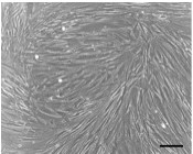 Fig. 1. Micrograph of HFL-I (Okano, N., 2017).
MRJP Prolonged the Life Span and Promoted the Proliferation of HFL-I Cells
Royal jelly (RJ), secreted from honeybees, has multiple known bioactivities, such as antioxidant and immunoregulatory effects. However, its specific impact on human cell aging required further exploration. Jiang's team studied the anti-senescence effects of major royal jelly proteins (MRJPs) on human embryonic lung fibroblast (HFL-I) cells. The findings showed that MRJPs significantly increased cell proliferation, reduced senescence and extended telomeres, suggesting that MRJPs can act as anti-senescence mediators in human cells.
Cellular morphological observations revealed that MRJPs affected the appearance of HFL-I cells. In medium A (Fig. 1a), most cells appeared flat with unclear boundaries, characteristic of senescence. Cells were often unattached and lost their spindle shape. Conversely, cells in medium C (Fig. 1c) and medium D (Fig. 1d) maintained a healthy shape and size with distinct boundaries. Cells in medium B (Fig. 1b) and medium E (Fig. 1e) exhibited signs of senescence. In comparison to medium A, PDL increased by 12.4%, 31.2%, 24.0% and 10.4% in media B, C, D, and E, respectively. Medium C cells exhibited the highest PDL, indicating that 0.2 mg/ml MRJPs (medium C) was the maximal dose to prolong the lifespan of HFL-I cells (Fig. 2a). By day 4, the proliferation (absorbance) of cells in media A, B, C, D, and E was 0.39, 0.40, 0.45, 0.44, and 0.42, respectively (Fig. 2b). RPA in media B, C, D and E increased by 1.0%, 11.9%, 11.1%, and 7.7% when compared with medium A.
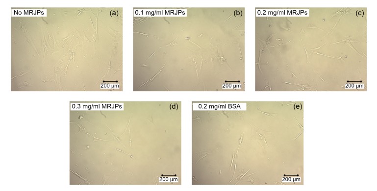 Fig. 1. Cellular morphology of HFL-I cells supplied with different concentrations of MRJPs or BSA (Jiang CM, Liu X, et al., 2018).
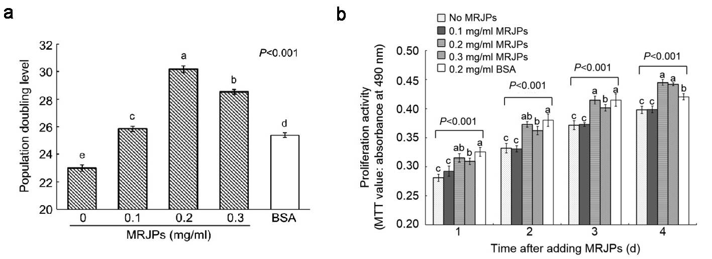 Fig. 2. (a) Lifespans of HFL-I cells supplied with different concentrations of MRJPs or BSA. (b) Proliferation effect of MRJPs after 4 d (Jiang CM, Liu X, et al., 2018).
In Vitro Reaction of Cells Derived from Human Normal Lung Tissues to Carbon-Ion Beam Irradiation
The translation of X-ray therapy data to CIRT is hindered by the unclear relative biological effectiveness (RBE) of carbon-ion beams in normal lung tissues. To overcome this, Okano utilized immortalized human small airway epithelial cells (iSAECs), human lung fibroblasts (HFL-1), and A549 lung cancer cells to compare the effects of X-ray and carbon-ion beam irradiation. He employed crystal violet staining (CVS) and clonogenic assays to generate survival curves, demonstrating the CVS assay's efficacy as an alternative for assessing the RBE of carbon-ion beams in non-clonogenic cells.
Using the CVS assay, irradiation of iSAECs and HFL-I cells with X-rays or carbon-ion beams decreased absorbance in a dose-dependent manner at low doses (Fig. 3A and B). However, high-dose irradiation caused cells to lose proliferative ability and show enlarged morphology (Fig. 3C and D), characteristic of senescence. Microscopic observation confirmed fewer cells after high-dose exposure than estimated by CVS assay, indicating an overestimation of the surviving fraction (Fig. 4). The induction of senescence in these cells was verified by increased SA-β-gal positivity 10 days post-irradiation with high doses. These findings indicate that the high staining intensity of the samples could be attributed to senescent cells with enlarged bodies that accumulated on the culture plates in response to high-dose irradiation, leading to the overestimation of the surviving fraction in the CVS assay.
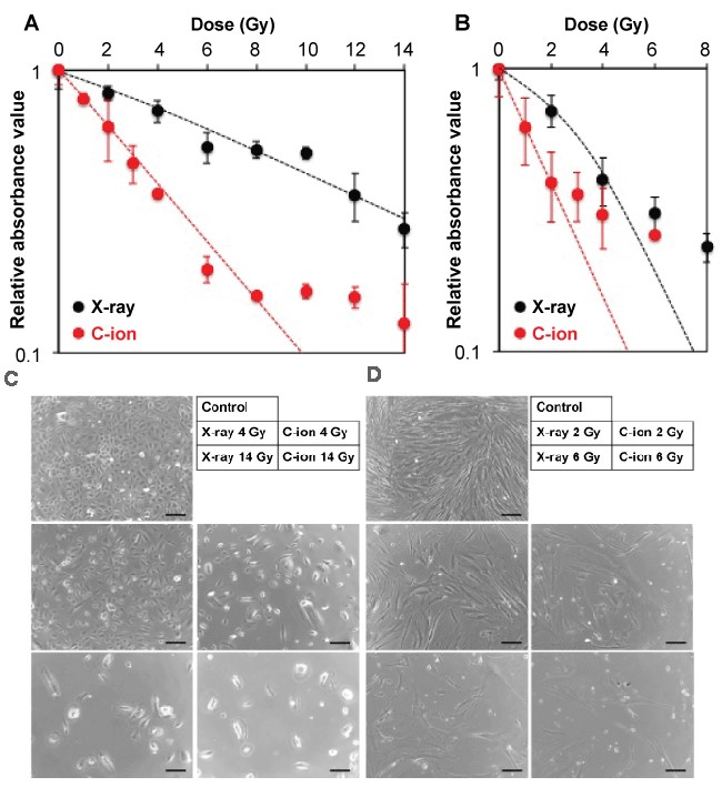 Fig. 3. (A-B) Sensitivity of iSAECs and HFL-I cells to X-ray and carbon-ion beam irradiation assessed by using the CVS assay. (C-D) Representative micrographs of iSAECs and HFL-I cells exposed to X-ray and carbon-ion beam irradiation (Okano, N., 2017).
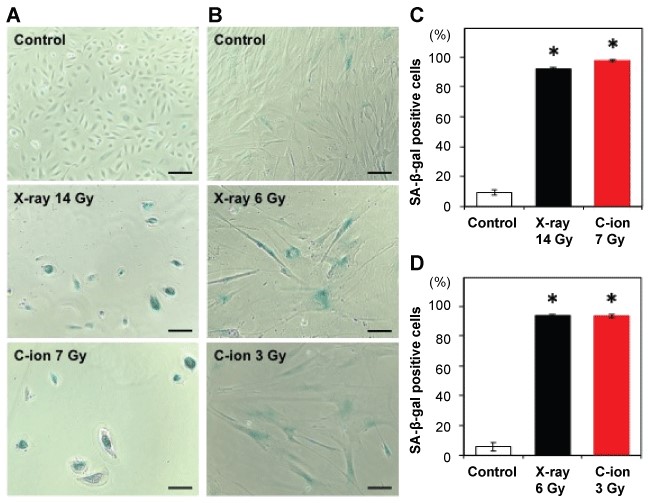 Fig. 4. Induction of senescence by X-ray and carbon-ion beam irradiation of iSAECs and HFL-I cells (Okano, N., 2017).
HFL-I is a fibroblast cell line that was isolated from the lung of a White, normal embryo. This cell line can be used in toxicology research.
Ask a Question
Average Rating: 5.0 | 5 Scientist has reviewed this product
High-quality & reliable
The cells performed very well in my experiments, a testament to their quality and reliability.
18 Mar 2023
Ease of use
After sales services
Value for money
High-quality & reliable
The cells performed very well in my experiments, a testament to their quality and reliability.
18 Mar 2023
Ease of use
After sales services
Value for money
High-quality & reliable
The cells performed very well in my experiments, a testament to their quality and reliability.
18 Mar 2023
Ease of use
After sales services
Value for money
High-quality & reliable
The cells performed very well in my experiments, a testament to their quality and reliability.
18 Mar 2023
Ease of use
After sales services
Value for money
High-quality & reliable
The cells performed very well in my experiments, a testament to their quality and reliability.
18 Mar 2023
Ease of use
After sales services
Value for money
Write your own review
- You May Also Need


