Transmission Electron Microscopy (TEM) Service
- Service Details
- Features
- Applications
- FAQ
- Explore Other Options
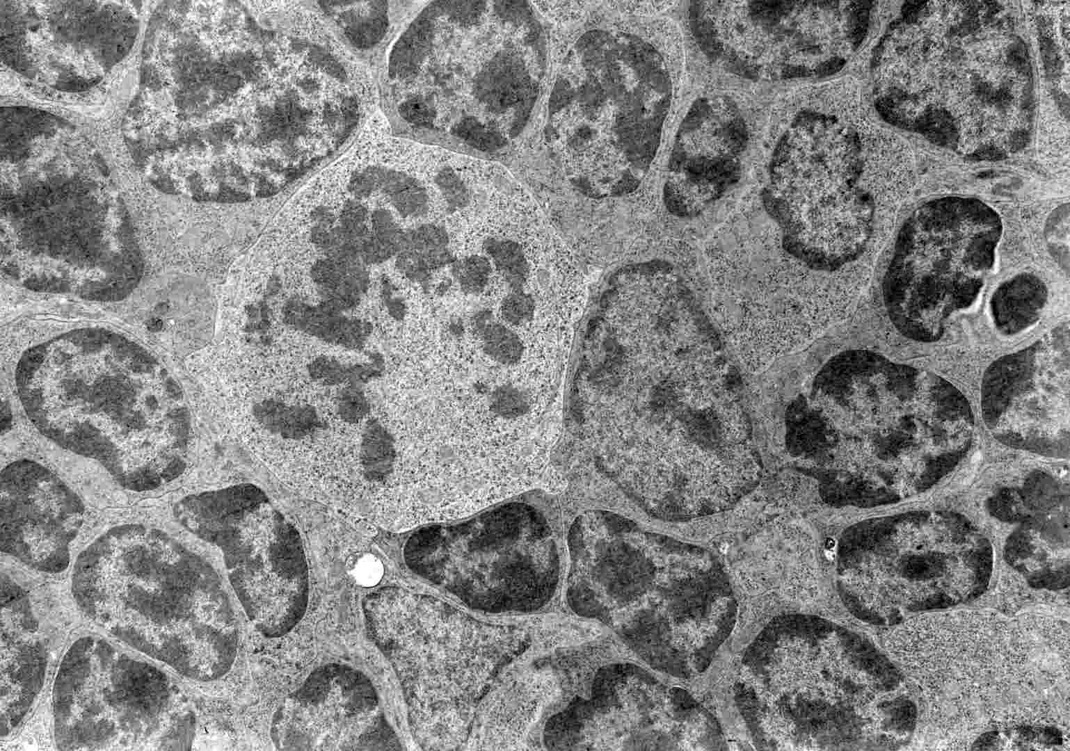
 TEM micrograph of a mitotic cell surrounded by interphase cells.
Transmission Electron Microscopy (TEM) stands out as a high-resolution imaging technology that employs high-energy electron beams to obtain atomic-level images of ultra-thin sample interiors. Scientists rely on TEM as an essential instrument for basic research and development across physics, chemistry, biology, and materials science because of its outstanding spatial resolution and flexible analytical features.
Creative Bioarray offers top-notch imaging and data solutions for scientific research and clinical diagnostics using cutting-edge TEM and STEM technologies. We provide analytical services for diverse biological materials including plant samples as well as animal tissues, bacterial samples and pathological specimens. Our seasoned experts deliver precise experimental design guidance for sample collection procedures along with preparation techniques and evaluation methods to achieve top analytical outcomes.
Creative Bioarray’s Transmission Electron Microscopy (TEM) Service
Available assays
Cryogenic Transmission Electron Microscopy (cryoTEM)
This method uses cryogenic techniques to maintain the sample's natural state and reduce radiation damage, making it perfect for examining the natural structures of biomolecules, organelles, and viruses. It offers high-resolution cryo-images that reveal intricate features and three-dimensional structures.
Negative Staining TEM
Enhances contrast in samples via negative staining, useful for examining the morphology and structure of nanoparticles, viruses, and biomolecules.
Particle Size Analysis (TEM, SEM)
This approach quantitatively analyzes the particle size distribution using TEM or SEM. TEM is ideal for nanoscale analysis, providing high-resolution images, while SEM is suitable for larger particles.
Immuno Transmission Electron Microscopy (immuno-TEM)
Combines immunolabeling with TEM, where specific antibodies bind to target molecules, marked by gold-labeled secondary antibodies, achieving precise localization and visualization of specific proteins, antigens, or antibodies.
Single-Particle Cryo-EM
This technology based on Cryo-TEM, reconstructing high-resolution three-dimensional structures by analyzing images of numerous individual particles, suitable for structural analysis of proteins, viruses, and nucleic acids.
Deliverables
- High-Resolution Images: Deliver clear, atomically detailed TEM images.
- Analysis Reports: Provide comprehensive analysis on morphology, structure, particle size, and elemental distribution.
- Data Files: Supply both raw image and analytical data files.
- Professional Consultation: Offer expert image interpretations and advice tailored to client needs.
Features

Advanced Equipment
Deploy cutting-edge transmission electron microscopy devices to achieve precise imaging and analysis results.

One-Stop Service
Offer complete solutions which include sample preparation and extend to image acquisition and data analysis to save clients both time and effort.

Experienced Expert Team
Our technical support team delivers professional assistance to guarantee successful implementation of complex projects.

Customized Solutions
Develop analytical plans that match the unique attributes of client samples to achieve the best possible outcomes.

Efficient Delivery
Our goal is to provide high-quality results within the shortest timeframe possible to keep client projects advancing smoothly.
What can You do with Our Transmission Electron Microscopy (TEM) Service?
- Structural Biology: 3D visualization of biomolecules such as proteins and nucleic acids enables researchers to study their functional mechanisms.
- Cell Line Characterization: Provides detailed views of cellular ultrastructure with specifics on organelle shapes and membrane positioning.
- Viral Vector Characterization: TEM technology examines viral vectors' morphology, structure, and purity during gene therapy and vaccine development to guarantee product safety and efficacy.
- Contaminant Detection: Identifies potential microbial or chemical contaminants in samples, including raw cell culture media and bioreactor harvests.
- Nanoparticle Characterization: Analyzes the size, shape, distribution, and surface characteristics of nanoparticles.
FAQ
1. What is TEM service?
The TEM service provides complete imaging and analytical support through transmission electron microscopy technology. High-energy electron beams penetrate ultra-thin samples to produce high-resolution images of their internal structures. Creative Bioarray provides TEM services which consist of experimental design guidance followed by sample collection and preparation and further image acquisition and analysis for applications in materials science, nanotechnology, and biology.
2. What should I consider for sample collection and fixation?
Samples should be fixed promptly after collection to preserve their native structure, typically using glutaraldehyde-containing fixatives like Modified Karnovsky's or McDowell-Trump fixative. If immediate fixation is not feasible, cutting the tissue into 1mm cubes is recommended to ensure thorough penetration of the fixative. The sample thickness should be less than 100 nanometers to facilitate electron beam transmission.
3. What is the TEM sample preparation process?
The TEM sample preparation process involves:
- Fixation: Chemically fix the sample to maintain its structure.
- Dehydration: Remove moisture using ethanol or acetone series.
- Embedding: Embed the sample in a resin block for sectioning.
- Sectioning: Use an ultramicrotome to cut the sample into thin slices (50-100 nanometers thick).
- Staining: Apply heavy metal stains (e.g., lead acetate or uranyl citrate) to enhance contrast.
- Mounting: Place sample slices on copper or nickel grids for electron microscope observation.
4. Can TEM technology be used to observe live cells?
Generally, TEM technology cannot be used to directly observe live cells because it requires the preparation of ultra-thin sections (approximately 100 nanometers thick), and the observation must be conducted in a high-vacuum environment, where live cells cannot survive. Additionally, the high-energy electron beam could cause radiation damage to cells. Thus, TEM is mainly used for observing the ultrastructure of fixed and dehydrated cells or tissues. However, the latest developments in cryo-electron microscopy aim to preserve some features of live cells.
5. What factors affect the resolution of TEM images?
The resolution of TEM images depends on multiple factors including the electron beam's accelerating voltage, sample thickness, electron source quality, sample preparation quality, lens system performance, and detector sensitivity.
Explore Other Options

