- You are here: Home
- Services
- Disease Models
- Pain Models
- Neuropathic Pain Models
- Spared Nerve Injury (SNI) Model
Services
-
Cell Services
- Cell Line Authentication
- Cell Surface Marker Validation Service
-
Cell Line Testing and Assays
- Toxicology Assay
- Drug-Resistant Cell Models
- Cell Viability Assays
- Cell Proliferation Assays
- Cell Migration Assays
- Soft Agar Colony Formation Assay Service
- SRB Assay
- Cell Apoptosis Assays
- Cell Cycle Assays
- Cell Angiogenesis Assays
- DNA/RNA Extraction
- Custom Cell & Tissue Lysate Service
- Cellular Phosphorylation Assays
- Stability Testing
- Sterility Testing
- Endotoxin Detection and Removal
- Phagocytosis Assays
- Cell-Based Screening and Profiling Services
- 3D-Based Services
- Custom Cell Services
- Cell-based LNP Evaluation
-
Stem Cell Research
- iPSC Generation
- iPSC Characterization
-
iPSC Differentiation
- Neural Stem Cells Differentiation Service from iPSC
- Astrocyte Differentiation Service from iPSC
- Retinal Pigment Epithelium (RPE) Differentiation Service from iPSC
- Cardiomyocyte Differentiation Service from iPSC
- T Cell, NK Cell Differentiation Service from iPSC
- Hepatocyte Differentiation Service from iPSC
- Beta Cell Differentiation Service from iPSC
- Brain Organoid Differentiation Service from iPSC
- Cardiac Organoid Differentiation Service from iPSC
- Kidney Organoid Differentiation Service from iPSC
- GABAnergic Neuron Differentiation Service from iPSC
- Undifferentiated iPSC Detection
- iPSC Gene Editing
- iPSC Expanding Service
- MSC Services
- Stem Cell Assay Development and Screening
- Cell Immortalization
-
ISH/FISH Services
- In Situ Hybridization (ISH) & RNAscope Service
- Fluorescent In Situ Hybridization
- FISH Probe Design, Synthesis and Testing Service
-
FISH Applications
- Multicolor FISH (M-FISH) Analysis
- Chromosome Analysis of ES and iPS Cells
- RNA FISH in Plant Service
- Mouse Model and PDX Analysis (FISH)
- Cell Transplantation Analysis (FISH)
- In Situ Detection of CAR-T Cells & Oncolytic Viruses
- CAR-T/CAR-NK Target Assessment Service (ISH)
- ImmunoFISH Analysis (FISH+IHC)
- Splice Variant Analysis (FISH)
- Telomere Length Analysis (Q-FISH)
- Telomere Length Analysis (qPCR assay)
- FISH Analysis of Microorganisms
- Neoplasms FISH Analysis
- CARD-FISH for Environmental Microorganisms (FISH)
- FISH Quality Control Services
- QuantiGene Plex Assay
- Circulating Tumor Cell (CTC) FISH
- mtRNA Analysis (FISH)
- In Situ Detection of Chemokines/Cytokines
- In Situ Detection of Virus
- Transgene Mapping (FISH)
- Transgene Mapping (Locus Amplification & Sequencing)
- Stable Cell Line Genetic Stability Testing
- Genetic Stability Testing (Locus Amplification & Sequencing + ddPCR)
- Clonality Analysis Service (FISH)
- Karyotyping (G-banded) Service
- Animal Chromosome Analysis (G-banded) Service
- I-FISH Service
- AAV Biodistribution Analysis (RNA ISH)
- Molecular Karyotyping (aCGH)
- Droplet Digital PCR (ddPCR) Service
- Digital ISH Image Quantification and Statistical Analysis
- SCE (Sister Chromatid Exchange) Analysis
- Biosample Services
- Histology Services
- Exosome Research Services
- In Vitro DMPK Services
-
In Vivo DMPK Services
- Pharmacokinetic and Toxicokinetic
- PK/PD Biomarker Analysis
- Bioavailability and Bioequivalence
- Bioanalytical Package
- Metabolite Profiling and Identification
- In Vivo Toxicity Study
- Mass Balance, Excretion and Expired Air Collection
- Administration Routes and Biofluid Sampling
- Quantitative Tissue Distribution
- Target Tissue Exposure
- In Vivo Blood-Brain-Barrier Assay
- Drug Toxicity Services
Spared Nerve Injury (SNI) Model
Creative Bioarray offers the spared nerve injury (SNI) model, a widely recognized experimental model, to our esteemed clients engaged in the study of neuropathic pain mechanisms or the preclinical evaluation of analgesic drug efficacy. Our team of experienced professionals ensures that all procedures are conducted with the highest standards of precision and ethical considerations, providing our clients with reliable and reproducible data to support your research. Whether you are aiming to uncover new insights into the molecular and cellular pathways involved in neuropathic pain or seeking to validate the effectiveness of your analgesic compounds, Creative Bioarray's SNI model is your trusted partner in advancing the frontiers of pain research.
Peripheral neuropathic pain is produced by multiple etiological factors that initiate a number of diverse mechanisms operating at different sites and at different times and expressed both within, and across different disease states. Unraveling the mechanisms involved requires laboratory animal models that replicate as far as possible, the different pathophysiological changes present in patients. The SNI model is a common experimental technique used in neuroscience research to induce neuropathic pain in animals, typically rodents. Researchers use the SNI model to study the mechanisms underlying neuropathic pain and to test potential treatments for chronic pain conditions.
Our Spared Nerve Injury (SNI) Model
- Available Animal
Rat
- Modeling Method
In the model group, after anesthetization, an approximately 2 cm incision is made to the upper edge of the left hind limb. Afterward, the muscle is separated gently, and the main trunk of the sciatic nerve and its 3 branches are exposed and bluntly separated. Then, the common peroneal nerve and tibial nerve are ligated with 4-0 silk sutures. Both nerves are severed at about 2 cm after ligation and the sural nerve was preserved. In the sham group, the sciatic nerve and its 3 branches are only exposed.
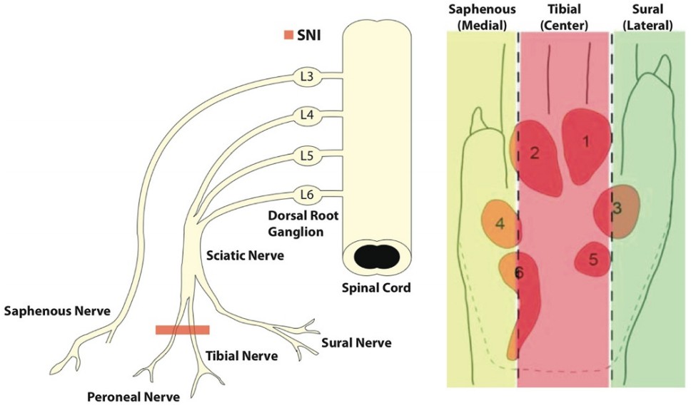 Fig. 1 The procedure of Spared Nerve Injury (SNI) Model. (Duraku et al. 2012)
- Endpoints
- Body weight
- Behavioral tests: Von Frey test, Hot plate test, etc.
- qPCR or Western blot
- Histology analysis
- Other customized endpoints
Example Data
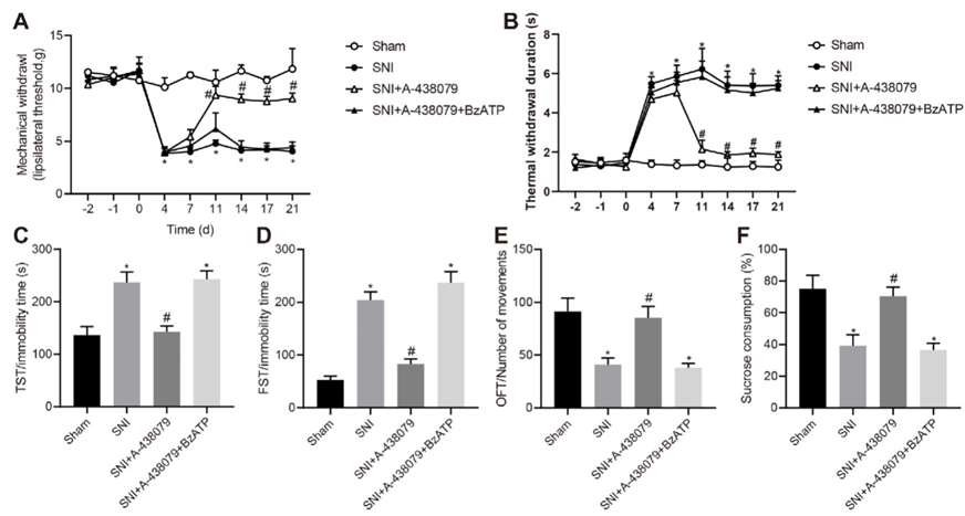 Fig. 2 Inhibition of P2X7R in the amygdala elevated the nociceptive threshold of MWT and TWD. (A) MWT was measured in rats of different groups with von Frey filaments. (B) TWD was measured in rats of different groups with von Frey filaments. (C) TST was performed in rats of different groups. (D) FST was carried out in rats of different groups. (E) OFT was conducted in rats of different groups. (F) SPT was performed in rats of different groups (21 days). (Hu et al. 2020)
Moreover, we also provide other neuropathic pain models that maybe you are interested in:
- Diabetes-Induced Neuropathic Pain Model
- Chemotherapy-Induced Neuropathic Pain Model
- Chronic Constriction Injury (CCI) Model
- Partial Sciatic Nerve Ligation (PSL/PSNL) Model
- Spinal Nerve Ligation (SNL) Model
Quotation and Ordering
Creative Bioarray is dedicated to providing stable disease models for our clients to accelerate your drug development. If you are interested in our services, please feel free to contact us at any time or submit an inquiry to us directly.
References
- Hu, X., et al. Inhibition of P2X7R in the amygdala ameliorates symptoms of neuropathic pain after spared nerve injury in rats. Brain, Behavior, and Immunity, 2020, 88: 507-514.
- Decosterd, I., Woolf, C.J. Spared nerve injury: an animal model of persistent peripheral neuropathic pain. Pain, 2000, 87(2): 149-158.
- Duraku, L.S., et al. Spatiotemporal dynamics of re-innervation and hyperinnervation patterns by uninjured CGRP fibers in the rat foot sole epidermis after nerve injury. Mol Pain. 2012; 8:61.
Explore Other Options
For research use only. Not for any other purpose.
