- You are here: Home
- Applications
- Neurological Disorder
- Alzheimer's Disease Modeling and Assays
Applications
-
Cell Services
- Cell Line Authentication
- Cell Surface Marker Validation Service
-
Cell Line Testing and Assays
- Toxicology Assay
- Drug-Resistant Cell Models
- Cell Viability Assays
- Cell Proliferation Assays
- Cell Migration Assays
- Soft Agar Colony Formation Assay Service
- SRB Assay
- Cell Apoptosis Assays
- Cell Cycle Assays
- Cell Angiogenesis Assays
- DNA/RNA Extraction
- Custom Cell & Tissue Lysate Service
- Cellular Phosphorylation Assays
- Stability Testing
- Sterility Testing
- Endotoxin Detection and Removal
- Phagocytosis Assays
- Cell-Based Screening and Profiling Services
- 3D-Based Services
- Custom Cell Services
- Cell-based LNP Evaluation
-
Stem Cell Research
- iPSC Generation
- iPSC Characterization
-
iPSC Differentiation
- Neural Stem Cells Differentiation Service from iPSC
- Astrocyte Differentiation Service from iPSC
- Retinal Pigment Epithelium (RPE) Differentiation Service from iPSC
- Cardiomyocyte Differentiation Service from iPSC
- T Cell, NK Cell Differentiation Service from iPSC
- Hepatocyte Differentiation Service from iPSC
- Beta Cell Differentiation Service from iPSC
- Brain Organoid Differentiation Service from iPSC
- Cardiac Organoid Differentiation Service from iPSC
- Kidney Organoid Differentiation Service from iPSC
- GABAnergic Neuron Differentiation Service from iPSC
- Undifferentiated iPSC Detection
- iPSC Gene Editing
- iPSC Expanding Service
- MSC Services
- Stem Cell Assay Development and Screening
- Cell Immortalization
-
ISH/FISH Services
- In Situ Hybridization (ISH) & RNAscope Service
- Fluorescent In Situ Hybridization
- FISH Probe Design, Synthesis and Testing Service
-
FISH Applications
- Multicolor FISH (M-FISH) Analysis
- Chromosome Analysis of ES and iPS Cells
- RNA FISH in Plant Service
- Mouse Model and PDX Analysis (FISH)
- Cell Transplantation Analysis (FISH)
- In Situ Detection of CAR-T Cells & Oncolytic Viruses
- CAR-T/CAR-NK Target Assessment Service (ISH)
- ImmunoFISH Analysis (FISH+IHC)
- Splice Variant Analysis (FISH)
- Telomere Length Analysis (Q-FISH)
- Telomere Length Analysis (qPCR assay)
- FISH Analysis of Microorganisms
- Neoplasms FISH Analysis
- CARD-FISH for Environmental Microorganisms (FISH)
- FISH Quality Control Services
- QuantiGene Plex Assay
- Circulating Tumor Cell (CTC) FISH
- mtRNA Analysis (FISH)
- In Situ Detection of Chemokines/Cytokines
- In Situ Detection of Virus
- Transgene Mapping (FISH)
- Transgene Mapping (Locus Amplification & Sequencing)
- Stable Cell Line Genetic Stability Testing
- Genetic Stability Testing (Locus Amplification & Sequencing + ddPCR)
- Clonality Analysis Service (FISH)
- Karyotyping (G-banded) Service
- Animal Chromosome Analysis (G-banded) Service
- I-FISH Service
- AAV Biodistribution Analysis (RNA ISH)
- Molecular Karyotyping (aCGH)
- Droplet Digital PCR (ddPCR) Service
- Digital ISH Image Quantification and Statistical Analysis
- SCE (Sister Chromatid Exchange) Analysis
- Biosample Services
- Histology Services
- Exosome Research Services
- In Vitro DMPK Services
-
In Vivo DMPK Services
- Pharmacokinetic and Toxicokinetic
- PK/PD Biomarker Analysis
- Bioavailability and Bioequivalence
- Bioanalytical Package
- Metabolite Profiling and Identification
- In Vivo Toxicity Study
- Mass Balance, Excretion and Expired Air Collection
- Administration Routes and Biofluid Sampling
- Quantitative Tissue Distribution
- Target Tissue Exposure
- In Vivo Blood-Brain-Barrier Assay
- Drug Toxicity Services
Alzheimer's Disease Modeling and Assays
Alzheimer's disease (AD) is the most common form of neurodegenerative disease, characterized by two aberrant features, the amyloid plaques and the neurofibrillary tangles. The initial pathophysiologic changes of AD were found in the hippocampal and entorhinal cortical region of the brain, which subsequently followed by disrupted learning and memory ability. AD progression was associated with neurodegeneration largely due to the accumulation of two protein aggregates: β-amyloid (Aβ) and tau. This cognitive and memory decline age-related disease causes suffering to the patient and their caregivers. Getting treatment or prevention of AD has attracted extensive attention worldwide. As a result, scientists are putting great efforts in understanding the mechanisms underlying the development of AD as well as treatment for AD.
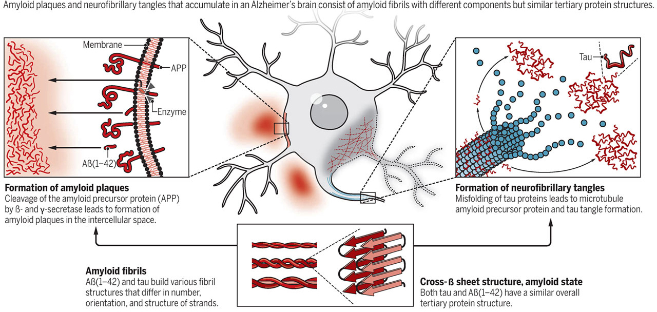
 Figure 1 Molecular characteristics of Alzheimer’s disease
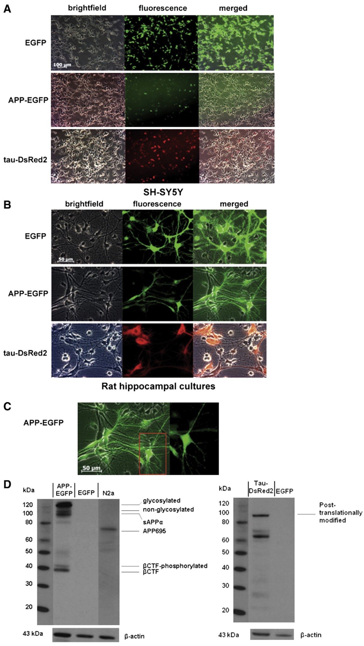
 Figure 2 Expression analysis of the transgenes in AD cell types.
The well-known neuropathogenic hallmarks of AD consist of aggregated amyloid beta (Aβ) and abnormal neurites. It is demonstrated that a dysregulated proteolytic processing of its precursor molecule, the Amyloid Precursor Protein (APP), can cause the accumulation of Aβ. Aβ can also cause caspases-mediated tau cleavage and hyperphosphorylation by activating specific kinases, thus promoting its aggregation, mis-localization and accumulation with consequent neurofibrillary tangles formation. On the other hand, Tau is a neuron specific microtubule-associated protein that regulates microtubule stability, tau dissociates from microtubules and forms insoluble aggregates called neurofibrillary tangles (NFTs). Both total tau (t-tau) and phosphorylated tau (p-tau) proteins are measured and associated with AD. Thus, Aβ peptide and tau exert crucial roles in neuronal loss or dysfunction, which would cause the increased reactive oxygen species (ROS) production, membrane damage, altered mitochondrial metabolism, abortive cell cycle events, and DNA damage/repair and inflammatory processes inevitably leading to neuronal dysfunction. Based on the above understanding, appropriate in vitro and in vivo models are deeply needed to mimic the pathogenesis and progression of AD, which could also work better with assays to study the drug candidates for Alzheimer's disease.
With extensive experience and state-of-the-art technologies, Creative Bioarray is offering Alzheimer's diseases modeling and assays to help our customers open the door for generating new insight into disease pathophysiology and improving the process of drug development.
Disease Modeling and assays available
In Vitro Model Service
- Various cell types (both cell lines and primary neuronal cultures; human cells or other species) are available for building Alzheimer's disease models according to your application.
- Our neurotoxicity assay, neuroprotective effect assay, and neurite outgrowth assay can cover most aspects for evaluation of new drugs.
- Customized assays and High-throughput screening are available.
- The final report will include the protocol of disease modeling and image analysis as well as statistical analysis.
In Vivo Model Service
- Diverse transgenic mouse models are developed and used to study gene function for AD pathologies or screen compounds or drugs for the treatment of AD.
- These transgenic mouse lines recapitulate different pathological and behavioral features in Alzheimer’s disease, and are available for drug development targeting related mechanisms.
- Endpoints are behavioral (memory loss) and histological.
Alzheimer's Disease Study Tools
- Screening preclinical drug candidates targeting Alzheimer's Disease using integrated assay tools.
- Evaluating changes in general cognitive function of AD models by behavioral tests.
- Revealing the three-dimensional anatomical structure and function of animal brain by neurological Imaging.
- Investigating neuronal properties in brain slices by patch-clamp.
Quotation and ordering
Contact us if you have any questions. Our customer service representatives are available 24hr a day.
References
- Pospich S, Raunser S. The molecular basis of Alzheimer's plaques. Science. 2017, 358(6359): 45.
- Stoppelkamp, S.; et al. In vitro modelling of Alzheimer's disease: degeneration and cell death induced by viral delivery of amyloid and tau. Experimental neurology. 2011, 229(2): 226-237.
Explore Other Options
For research use only. Not for any other purpose.

