- You are here: Home
- Disease Models
- Cardiovascular Disease Models
- Heart Failure with Preserved Ejection Fraction (HFpEF) Model
Disease Models
- Oncology Models
-
Inflammation & Autoimmune Disease Models
- Rheumatoid Arthritis Models
- Glomerulonephritis Models
- Multiple Sclerosis (MS) Models
- Ocular Inflammation Models
- Sjögren's Syndrome Model
- LPS-induced Acute Lung Injury Model
- Peritonitis Models
- Passive Cutaneous Anaphylaxis Model
- Delayed-Type Hypersensitivity (DTH) Models
- Inflammatory Bowel Disease Models
- Systemic Lupus Erythematosus Animal Models
- Asthma Model
- Sepsis Model
- Psoriasis Model
- Atopic Dermatitis (AD) Model
- Scleroderma Model
- Gouty Arthritis Model
- Carrageenan-Induced Air Pouch Synovitis Model
- Carrageenan-Induced Paw Edema Model
- Experimental Autoimmune Myasthenia Gravis (EAMG) Model
-
Cardiovascular Disease Models
- Surgical Models
- Animal Models of Hypertension
- Venous Thrombosis Model
- Atherosclerosis model
- Cardiac Arrhythmia Model
- Hyperlipoidemia Model
- Doxorubicin-induced Heart Failure Model
- Isoproterenol-induced Heart Failure Model
- Arterial Thrombosis Model
- Pulmonary Arterial Hypertension (PAH) Models
- Heart Failure with Preserved Ejection Fraction (HFpEF) Model
-
Neurological Disease Models
- Alzheimer's Disease Modeling and Assays
- Seizure Models
- Parkinson's Disease Models
- Ischemic Stroke Models
- Acute Spinal Cord Injury (ASCI) Model
- Traumatic Brain Injury (TBI) Model
- Hypoxic-Ischemic Encephalopathy (HIE) Model
- Tourette Syndrome (TS) Model
- Amyotrophic Lateral Sclerosis (ALS) Model
- Huntington's Disease (HD) Model
- Intracerebral hemorrhage (ICH) Models
- Pain Models
- Metabolic Disease Models
- Liver Disease Models
- Rare Disease Models
- Respiratory Disease Models
- Digestive Disease Models
-
Urology Disease Models
- Cisplatin-induced Nephrotoxicity Model
- Unilateral Ureteral Obstruction Model
- 5/6 Nephrectomy Model
- Renal Ischemia-Reperfusion Injury (RIRI) Model
- Diabetic Nephropathy (DN) Models
- Passive Heymann Nephritis (PHN) Model
- Adenine-Induced Chronic Kidney Disease (CKD) Model
- Kidney Stone Model
- Doxorubicin-Induced Nephropathy Model
- Orthopedic Disease Models
- Ocular Disease Models
- Skin Disease Models
- Infectious Disease Models
Heart Failure with Preserved Ejection Fraction (HFpEF) Model
At Creative Bioarray, we have successfully developed a robust animal model of heart failure with preserved ejection fraction (HFpEF) in mice. This model is specifically designed to evaluate the efficacy of your drug candidates by closely replicating the complex pathophysiology of HFpEF. By providing a reliable platform for assessing the therapeutic potential of various compounds, we support your drug development efforts with precision and reproducibility.
HFpEF is characterized by a left ventricular ejection fraction (LVEF) of 50% or higher, impaired diastolic function, and elevated levels of natriuretic peptides, all while presenting with symptoms and signs typical of heart failure. As HFpEF increasingly becomes the predominant form of heart failure, the therapeutic options available for this patient population remain limited. Understanding the heterogeneity of HFpEF is essential to developing effective treatments, making the use of animal and cellular models critical for exploring the underlying pathobiology and identifying new therapeutic agents.
Our Heart Failure with Preserved Ejection Fraction (HFpEF) Model
Animal Species
Mouse
Modeling Method
Animals are subjected 60% high fat diet and drinking water with L-NAME.
Endpoints
- Body weight
- Heart weight
- Exercise exhaustion test
- Echocardiography
- Histology analysis
- Biochemical analysis
- qPCR or Western blot
- Other customized endpoints according to your specific needs
Example Data
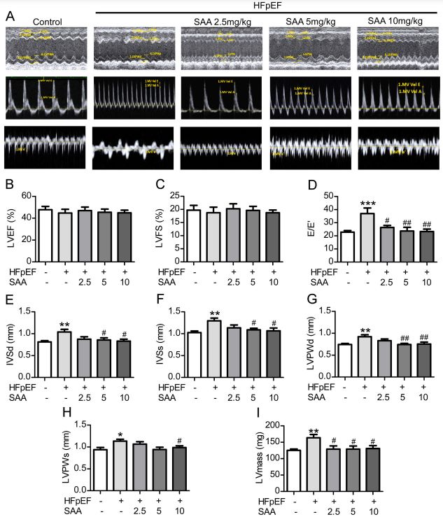 Fig. 1 SAA administration ameliorates left ventricular diastolic dysfunction in HFpEF mice. (A) Representative images conventional echocardiography and doppler imaging. (B) LVEF, left ventricular ejection fraction. (C) LVFS, left ventricular fractional shortening. (D) E/E', ratio between mitral E wave and E' wave. (E) IVSd, end-diastolic interventricular septal wall thickness. (F) IVSs, end-systolic interventricular septal wall thickness. (G) LVPWd, left ventricular end-diastolic posterior wall. (H) LVPWs, left ventricular end-systolic posterior wall. (I) LV mass, left ventricular mass. (Dawuti et al., 2023)
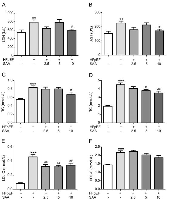 Fig. 2 SAA regulated heart injury and lipid metabolism disorder of HFpEF mice. (A) Lactate dehydrogenase (LDH). (B) Aspartate aminotransferase (AST). (C) Triglyceride (TG). (D) Total cholesterol (TC). (E) Low density lipoprotein cholesterol (LDL-C). (F) High density lipoprotein cholesterol (HDL-C). (Dawuti et al., 2023)
Quotation and Ordering
Creative Bioarray takes pride in its team of highly skilled experts and scientists who specialize in the development of cardiovascular disease models, model selection, and the execution of preclinical experiments. We are thrilled to leverage our cutting-edge technology and deep expertise to elevate our clients' research initiatives and increase their value. If you find our services appealing, we invite you to contact us whenever it suits you best or to send us an inquiry directly.
Reference
- Dawuti, A., et al. Salvianolic acid A alleviates heart failure with preserved ejection fraction via regulating TLR/Myd88/TRAF/NF-κB and p38MAPK/CREB signaling pathways. Biomedicine & Pharmacotherapy, 2023, 168: 115837.
For research use only. Not for any other purpose.
