QuantiGene Plex Assay - High-Throughput Gene Expression Analysis
- Service Details
- Features
- FAQ
- Explore Other Options
QuantiGene Plex Assay is an efficient multiplex gene expression quantification analysis technique that utilizes branched DNA (bDNA) technology for signal amplification. This method allows for the simultaneous quantification of up to 80 target genes in a single well of a 96-well or 384-well plate. This hybridization-based technology uses specific probes to bind with target RNA transcripts, forming complex branched structures through signal amplification of the probes, significantly enhancing detection sensitivity. The operational procedure of the QuantiGene Plex Assay is similar to that of an ELISA, requiring no RNA purification, cDNA synthesis, or PCR amplification, thereby simplifying the experimental steps.
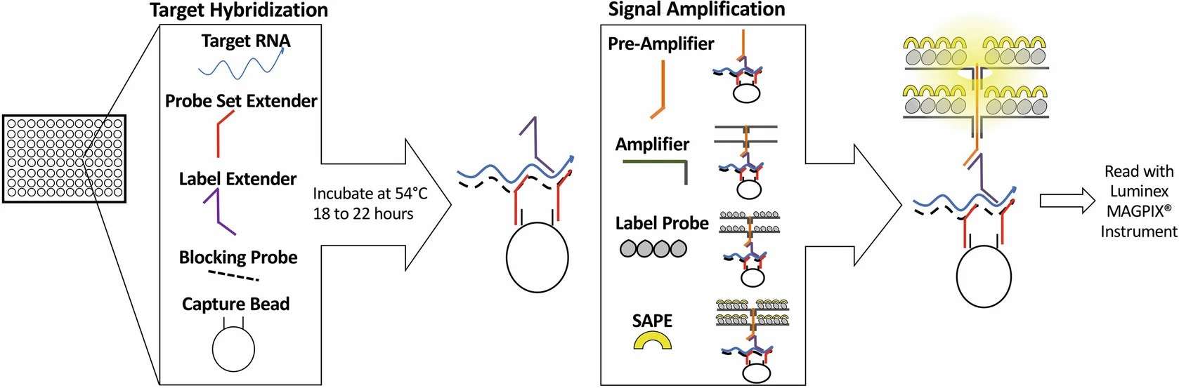 Fig. 1. Diagram of the QuantiGene Plex assay protocol (Dang T, Wang J, et al., 2022).
Applications
- Drug candidates screening
- Biomarker verification
- siRNA knockdown efficiency detection
- miRNA profiling
- Microarray validation
- Predictive toxicology
- Translocations and fusion genes detection.
QuantiGene Plex Assay available for many species, shown below:
- Human
- Cow
- HPV
- Atlantic salmon
- Mouse/Rat
- Chicken
- Horse
- Hamster
- Dog
- Monkey
- Rabbit
- House sparrow
- Pig
- Guniea pig
- Zebra fish
- Vibrio harvey
Creative Bioarray's QuantiGene Plex Assay Workflow
1
Sample Preparation
Begin by lysing the samples to release and stabilize the RNA. This process accommodates a broad range of samples, including cells, fresh/frozen tissue (plants/animal), FFPE tissues, blood, or purified RNA.
2
Target Hybridization
Incubate the samples overnight in multi-well plates (96- or 384-well) with specific probe sets that consist of capture extenders, label extenders, and blocking probes designed to hybridize with the target mRNA.
3
Signal Amplification
Employ bDNA technology to enhance the signal. The signal amplification process involves attaching specific probes to the mRNA target sequence. The target sequence, once hybridized, is fixed onto a bead through a capture probe, ensuring signal specificity. Amplification is achieved by adding a pre-amplifier that overlaps with a label extender, followed by multiple amplifiers that create a branched DNA structure. This structure features sites for biotinylated label probes, greatly enhancing the signal.
4
Detection
The biotinylated bDNA binds streptavidin conjugated R-phycoerythrin (SAPE), which results in a fluorescent signal proportional to the amount of target RNA. Then the signal detected by excitation with a laser using a Luminex instrument.
5
Data Analysis
Analyze the data through a cloud-based application, which allows for advanced analysis and visualization of the results from any location.
Features
| Feature | Benefit |
| High Multiplexing Capability | Detect up to 80 targets in a single well without cross-reactivity issues. |
| Simple Workflow | No need for RNA purification or PCR, reducing time and errors. |
| Resistance to Inhibitors | Works with challenging samples like FFPE tissues and whole blood. |
| Standardized Compatibility | Compatible with Luminex 200, FlexMap 3D, and xMAP Intelliflex platforms. |
FAQ
1. What sample types does the QuantiGene Plex Assay support?
Answer: Supports various sample types, including FFPE tissue, fresh or frozen tissue, whole blood, cultured cells, bacteria, and viruses, etc.
2. If I have specific genes that need to be tested, can you provide customized services?
Answer: Yes, we offer customized testing panel services, allowing clients to select specific genes for combination testing based on their needs. At the same time, we have a large verified gene database available for customers to choose from.
3. Which species' genes can be detected?
Currently, the QuantiGene Plex Assay technology can be widely applied to gene expression detection in common model organisms such as humans, mice, and rats. For gene detection needs in other species, we will evaluate and customize development based on specific circumstances.
4. What types of genes can be tested?
Our services can cover a wide range of gene types, including but not limited to cancer markers, apoptosis-related genes, inflammatory factors, and more.
Quotation and Ordering
For more detailed information, please feel free to contact us or directly sent us an inquiry. We look forward to cooperating with you in the future.
Reference
- Dang T, Wang J, et al. QuantiGene Plex Assay: A Method for High-Throughput Multiplex Citrus Viroid Detection and Identification. Methods Mol Biol. 2022. 2316:243-250.
Explore Other Options
