- You are here: Home
- Services
- Cell Services
- Cell Line Testing and Assays
- Phagocytosis Assays
Services
-
Cell Services
- Cell Line Authentication
- Cell Surface Marker Validation Service
-
Cell Line Testing and Assays
- Toxicology Assay
- Drug-Resistant Cell Models
- Cell Viability Assays
- Cell Proliferation Assays
- Cell Migration Assays
- Soft Agar Colony Formation Assay Service
- SRB Assay
- Cell Apoptosis Assays
- Cell Cycle Assays
- Cell Angiogenesis Assays
- DNA/RNA Extraction
- Custom Cell & Tissue Lysate Service
- Cellular Phosphorylation Assays
- Stability Testing
- Sterility Testing
- Endotoxin Detection and Removal
- Phagocytosis Assays
- Cell-Based Screening and Profiling Services
- 3D-Based Services
- Custom Cell Services
- Cell-based LNP Evaluation
-
Stem Cell Research
- iPSC Generation
- iPSC Characterization
-
iPSC Differentiation
- Neural Stem Cells Differentiation Service from iPSC
- Astrocyte Differentiation Service from iPSC
- Retinal Pigment Epithelium (RPE) Differentiation Service from iPSC
- Cardiomyocyte Differentiation Service from iPSC
- T Cell, NK Cell Differentiation Service from iPSC
- Hepatocyte Differentiation Service from iPSC
- Beta Cell Differentiation Service from iPSC
- Brain Organoid Differentiation Service from iPSC
- Cardiac Organoid Differentiation Service from iPSC
- Kidney Organoid Differentiation Service from iPSC
- GABAnergic Neuron Differentiation Service from iPSC
- Undifferentiated iPSC Detection
- iPSC Gene Editing
- iPSC Expanding Service
- MSC Services
- Stem Cell Assay Development and Screening
- Cell Immortalization
-
ISH/FISH Services
- In Situ Hybridization (ISH) & RNAscope Service
- Fluorescent In Situ Hybridization
- FISH Probe Design, Synthesis and Testing Service
-
FISH Applications
- Multicolor FISH (M-FISH) Analysis
- Chromosome Analysis of ES and iPS Cells
- RNA FISH in Plant Service
- Mouse Model and PDX Analysis (FISH)
- Cell Transplantation Analysis (FISH)
- In Situ Detection of CAR-T Cells & Oncolytic Viruses
- CAR-T/CAR-NK Target Assessment Service (ISH)
- ImmunoFISH Analysis (FISH+IHC)
- Splice Variant Analysis (FISH)
- Telomere Length Analysis (Q-FISH)
- Telomere Length Analysis (qPCR assay)
- FISH Analysis of Microorganisms
- Neoplasms FISH Analysis
- CARD-FISH for Environmental Microorganisms (FISH)
- FISH Quality Control Services
- QuantiGene Plex Assay
- Circulating Tumor Cell (CTC) FISH
- mtRNA Analysis (FISH)
- In Situ Detection of Chemokines/Cytokines
- In Situ Detection of Virus
- Transgene Mapping (FISH)
- Transgene Mapping (Locus Amplification & Sequencing)
- Stable Cell Line Genetic Stability Testing
- Genetic Stability Testing (Locus Amplification & Sequencing + ddPCR)
- Clonality Analysis Service (FISH)
- Karyotyping (G-banded) Service
- Animal Chromosome Analysis (G-banded) Service
- I-FISH Service
- AAV Biodistribution Analysis (RNA ISH)
- Molecular Karyotyping (aCGH)
- Droplet Digital PCR (ddPCR) Service
- Digital ISH Image Quantification and Statistical Analysis
- SCE (Sister Chromatid Exchange) Analysis
- Biosample Services
- Histology Services
- Exosome Research Services
- In Vitro DMPK Services
-
In Vivo DMPK Services
- Pharmacokinetic and Toxicokinetic
- PK/PD Biomarker Analysis
- Bioavailability and Bioequivalence
- Bioanalytical Package
- Metabolite Profiling and Identification
- In Vivo Toxicity Study
- Mass Balance, Excretion and Expired Air Collection
- Administration Routes and Biofluid Sampling
- Quantitative Tissue Distribution
- Target Tissue Exposure
- In Vivo Blood-Brain-Barrier Assay
- Drug Toxicity Services
Phagocytosis Assays
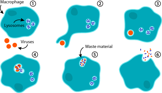
Phagocytosis is a specific form of endocytosis where cells internalize solid matter, thereby eliminating cellular debris and pathogens. Although most cells are capable of phagocytosis, the phagocytes in the immune system including neutrophils, macrophages and immature dendritic cells, play a key role in this process. In these cells, phagocytosis is a mechanism by which micro-organisms can be contained, killed and processed for antigen presentation and represents an important facet of the innate immune response, and plays an essential role in initiating the adaptive immune response.
There are many types of phagocytosis assays. By quantifying the engulfment of the certain substrate, we can obtain the phagocytic efficiency and then evaluate the effect of antibodies, vaccine, ligands, and the like.
Traditionally, erythrocytes are commonly used in phagocytosis assays. For FcR-mediated phagocytosis, erythrocytes are first opsonized with serum or IgG before added to phagocytes. After removal of non-phagocytic erythrocytes, engulfed erythrocytes are quantitated.
In contrary to conventional phagocytosis assays, the flow assay does not involve subjective manual counting of erythrocytes. This format provides a quantitative, high-throughput method to accurately measure phagocytosis.

Based on the superior expertise and extensive experiences, Creative Bioarray has developed a systematic approach to assess phagocytosis. These approaches are designed to help you analyze the function of specialized immune cells, evaluate the effects of antibodies and vaccine, as well as screen for TLR ligands, phagocytic activators and inhibitors.
Our Capabilities
- Phagocytosis assay - E. coli Substrate
- Phagocytosis assay - Red Blood Cell Substrate
- Phagocytosis assay - Zymosan Substrate
Features of Our Services
- Fully quantify phagocytosis with no manual cell counting
- High-throughput 96-well format
- Convenient quantitation in a standard microplate reader
If you are interested in our services, please feel free to contact us and our team will get in touch with you as soon as possible.
Explore Other Options
For research use only. Not for any other purpose.

