3D Tumor Spheroid Assay
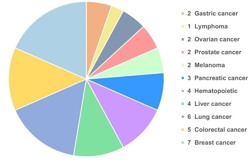
Creative Bioarray has validated multiple 3D tumor spheroid models that can be applied in drug screening and scientific research. Compared to traditional 2D models, 3D tumor spheroid models are more biomimetic, efficient, and cost-effective.
 Figure 1. Creative Bioarray has validated multiple 3D tumor spheroid models.
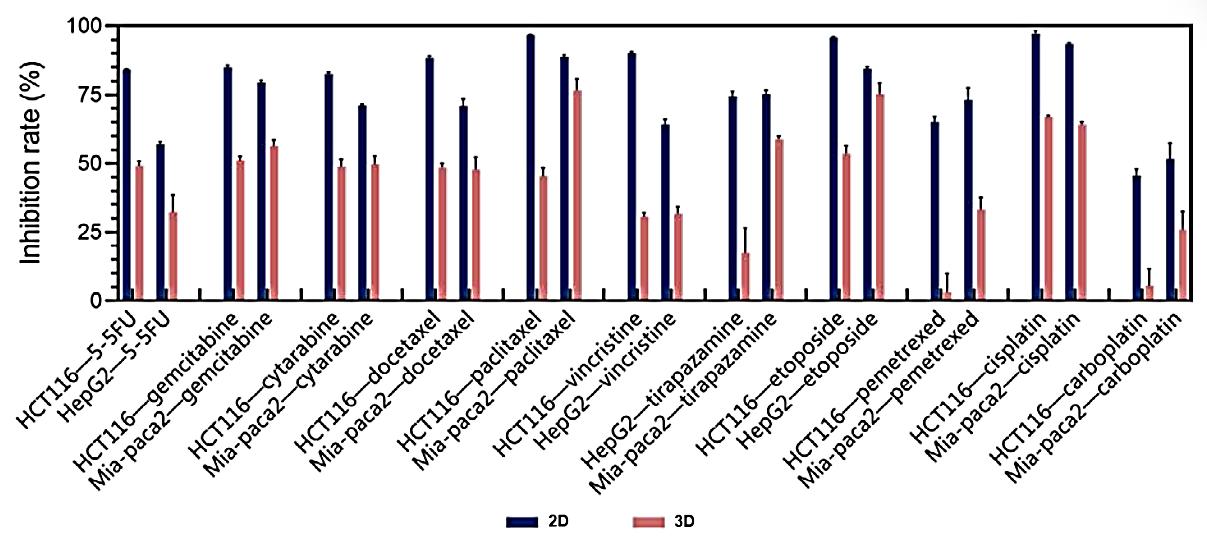
To evaluate the differences in sensitivity and specificity between 2D and 3D tumor cell models, we performed tests using commercially available chemotherapy drugs on multiple cell lines, including gastric cancer, ovarian cancer, prostate cancer, pancreatic cancer, liver cancer, lung cancer, colon cancer, breast cancer, and others. Additionally, we conducted tests using targeted and non-targeted drugs on specific cell lines with known targets, such as HCC827 (EGFR) and BT474 (Her2) cells. The results demonstrated a clear advantage in drug screening outcomes when utilizing 3D tumor models over 2D models.
Highlights
- 3D tumor spheroid models are more resistant to chemotherapy drugs.
The results of initial screening of chemotherapeutic drugs in tumor cell lines showed that the drug resistance of 3D tumor cells was generally stronger than that of traditional 2D model (Figure 2).
 Figure 2. Comparison of cell inhibition rates between 3D and 2D tumors models.
- 3D tumor spheroid models are more sensibility and specificity to targeted drugs.
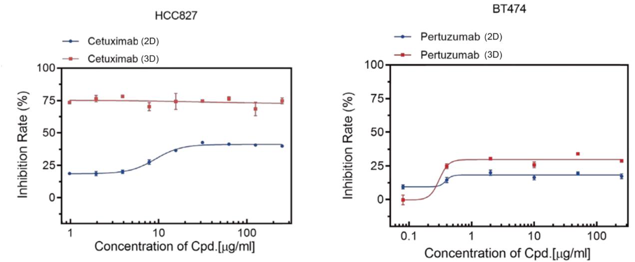
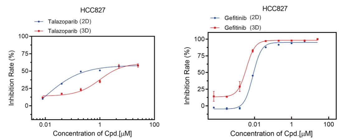
2D models will suffer from false positive screening results leading to non-target drugs cannot be excluded. HCC827 (EGFR) and BT474 (Her2) 3D tumor spheroid models exhibited a more sensitive growth inhibition response to Cetuximab and Pertuzumab, respectively (Figure 3). Meanwhile, HCC827 (EGFR) 3D tumor spheroid models showed higher resistance to the Talazoparib (Figure 4).
 Figure 3. Comparison of inhibition curves of the macromolecular drugs on different HCC827 and BT474 models.
 Figure 4. Comparison of inhibition curves of the Talazoparib and Gefitinib on different HCC827 models.
Creative Bioarray is committed to providing high-quality services and products to support cancer research and drug discovery. Our 3D Tumor Spheroid Assay is a powerful tool for evaluating the efficacy of potential anti-cancer drugs. Contact us today to learn more about our 3D Tumor Spheroid Assay and how it can benefit your research projects.
Applications
- High-throughput screening of active compounds
- Drug efficacy evaluation
Process of Analyzing drug response in 3D spheroid models

Study Examples:
 Figure 4: The anti-cancer drug test was performed in HCT116 spheroids. (B) HCT116 spheroid formation using the 3D culture system (left, 20×) and the spheroids collected from gels (right, 40×). (C) After 4 days of OXA treatment, spheroid cell viability was examined using Cell Titer-Glo® 3D Cell Viability Assay. [1]
Reference
- Yang, Cian-Ru et al. "A Novel 3D Culture Scaffold to Shorten Development Time for Multicellular Tumor Spheroids." International journal of molecular sciences vol. 23,22 13962. 12 Nov. 2022, doi:10.3390/ijms232213962
Explore Other Options