- You are here: Home
- Applications
- Neurological Disorder
- Parkinson's Disease Modeling and Assays
Applications
-
Cell Services
- Cell Line Authentication
- Cell Surface Marker Validation Service
-
Cell Line Testing and Assays
- Toxicology Assay
- Drug-Resistant Cell Models
- Cell Viability Assays
- Cell Proliferation Assays
- Cell Migration Assays
- Soft Agar Colony Formation Assay Service
- SRB Assay
- Cell Apoptosis Assays
- Cell Cycle Assays
- Cell Angiogenesis Assays
- DNA/RNA Extraction
- Custom Cell & Tissue Lysate Service
- Cellular Phosphorylation Assays
- Stability Testing
- Sterility Testing
- Endotoxin Detection and Removal
- Phagocytosis Assays
- Cell-Based Screening and Profiling Services
- 3D-Based Services
- Custom Cell Services
- Cell-based LNP Evaluation
-
Stem Cell Research
- iPSC Generation
- iPSC Characterization
-
iPSC Differentiation
- Neural Stem Cells Differentiation Service from iPSC
- Astrocyte Differentiation Service from iPSC
- Retinal Pigment Epithelium (RPE) Differentiation Service from iPSC
- Cardiomyocyte Differentiation Service from iPSC
- T Cell, NK Cell Differentiation Service from iPSC
- Hepatocyte Differentiation Service from iPSC
- Beta Cell Differentiation Service from iPSC
- Brain Organoid Differentiation Service from iPSC
- Cardiac Organoid Differentiation Service from iPSC
- Kidney Organoid Differentiation Service from iPSC
- GABAnergic Neuron Differentiation Service from iPSC
- Undifferentiated iPSC Detection
- iPSC Gene Editing
- iPSC Expanding Service
- MSC Services
- Stem Cell Assay Development and Screening
- Cell Immortalization
-
ISH/FISH Services
- In Situ Hybridization (ISH) & RNAscope Service
- Fluorescent In Situ Hybridization
- FISH Probe Design, Synthesis and Testing Service
-
FISH Applications
- Multicolor FISH (M-FISH) Analysis
- Chromosome Analysis of ES and iPS Cells
- RNA FISH in Plant Service
- Mouse Model and PDX Analysis (FISH)
- Cell Transplantation Analysis (FISH)
- In Situ Detection of CAR-T Cells & Oncolytic Viruses
- CAR-T/CAR-NK Target Assessment Service (ISH)
- ImmunoFISH Analysis (FISH+IHC)
- Splice Variant Analysis (FISH)
- Telomere Length Analysis (Q-FISH)
- Telomere Length Analysis (qPCR assay)
- FISH Analysis of Microorganisms
- Neoplasms FISH Analysis
- CARD-FISH for Environmental Microorganisms (FISH)
- FISH Quality Control Services
- QuantiGene Plex Assay
- Circulating Tumor Cell (CTC) FISH
- mtRNA Analysis (FISH)
- In Situ Detection of Chemokines/Cytokines
- In Situ Detection of Virus
- Transgene Mapping (FISH)
- Transgene Mapping (Locus Amplification & Sequencing)
- Stable Cell Line Genetic Stability Testing
- Genetic Stability Testing (Locus Amplification & Sequencing + ddPCR)
- Clonality Analysis Service (FISH)
- Karyotyping (G-banded) Service
- Animal Chromosome Analysis (G-banded) Service
- I-FISH Service
- AAV Biodistribution Analysis (RNA ISH)
- Molecular Karyotyping (aCGH)
- Droplet Digital PCR (ddPCR) Service
- Digital ISH Image Quantification and Statistical Analysis
- SCE (Sister Chromatid Exchange) Analysis
- Biosample Services
- Histology Services
- Exosome Research Services
- In Vitro DMPK Services
-
In Vivo DMPK Services
- Pharmacokinetic and Toxicokinetic
- PK/PD Biomarker Analysis
- Bioavailability and Bioequivalence
- Bioanalytical Package
- Metabolite Profiling and Identification
- In Vivo Toxicity Study
- Mass Balance, Excretion and Expired Air Collection
- Administration Routes and Biofluid Sampling
- Quantitative Tissue Distribution
- Target Tissue Exposure
- In Vivo Blood-Brain-Barrier Assay
- Drug Toxicity Services
Parkinson's Disease Modeling and Assays
Parkinson's disease (PD) is a progressive neurological disorder defined by a characteristic clinical syndrome of bradykinesia, tremor, rigidity, and postural instability. PD is the second most common neurodegenerative disease after Alzheimer’s disease, and the most frequent subcortical degenerative disease. It affects 1-2% of the people older than 60 years. The socio-economic cost of PD is high. In the United States, the cost per patient per year is around $10,000, with a total economic burden of $23 billion.
Neuronal loss in the substantia nigra pars compacta (SNc) and the subsequent loss of striatal dopamine content are regarded responsible for the classical motor features of PD. The misfolded α-synuclein is a major component of pathological perspective in neuronal loss. The toxic α-synuclein accumulation impairs the functions of mitochondria, lysosomes, and endoplasmic reticulum, and interferes with microtubular transport. Oxidative stress remains a cornerstone of the concepts underlying the loss of dopaminergic neurons in PD, which involves mitochondria and endoplasmic reticulum. Free radical production results in increased chemical and enzymatic oxidation of dopamine, which leads to the production of toxins such as 6-hydroxydopamine (6-OHDA). Besides, altered accumulation of iron in SNc, changes in calcium channel activity, altered proteolysis (proteasomal and lysosomal), changes in α-synuclein aggregation, and the presence of mutant proteins are all examples of how oxidative stress might be induced in PD.
Currently drug treatment provides only symptomatic relief. There is a great demand to explore how knowledge of the pathogenic processes and the use of experimental models of PD interlink to assist in the searching for neuroprotective/neurorestorative treatments.
With extensive experience and state-of-the-art technologies, Creative Bioarray is offering Parkinson's disease modeling and assays to help our customer generating new insight into disease pathophysiology and accelerating the process of drug development.
Advantages
- Various cells types (both cell lines and primary neuronal cultures; human cells or other species) are available for building Parkinson's disease models according to your application.
- Multiple evaluation aspects for new drugs.
- Customized assays and High-throughput screening are available on request.
Disease modeling and assays available
Creative Bioarray offers the development of custom designed in vitro models by using primary neuron cultures, cell lines, iPS cells with genetic modifications, which are phenotypically closer to the neuronal network and mimic PD pathology. We could also offer a wide range of assays that enable more accurate prediction of patient response to pharmacotherapy:
- Neuronal viability assays (including cell numbers, neurite outgrowth, and apoptosis)
- Neuro imaging for Cellular Morphology
- Oxidative Stress test
- Calcium signaling assay
- Mitochondrial dysfunction assay
We provide this powerful and versatile tool for PD therapy as well as basic research to help our customer understand more about the underlying mechanisms in the progression of PD.
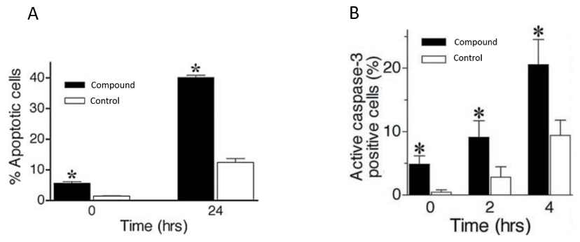
 Figure 1. Oxidative stress (exposure to H2O2 treatment) can increase apoptosis and caspase-3 activation in human neuroblastoma cells.
Quotation and ordering
Our customer service representatives are available 24hr a day!
References
- Dickson, Dennis W. Parkinson's disease and parkinsonism: neuropathology. Cold Spring Harbor perspectives in medicine. 2012, 2.8: a009258.
- Dexter, D., and Peter J. Parkinson disease: from pathology to molecular disease mechanisms. Free Radical Biology and Medicine. 2013, 62: 132-144.
- Sherer, T., et al. An in vitro model of Parkinson's disease: linking mitochondrial impairment to altered α-synuclein metabolism and oxidative damage. Journal of Neuroscience. 2002, 22.16: 7006-7015.
Explore Other Options
For research use only. Not for any other purpose.

