Single-Particle Cryo-EM
Single-Particle cryo-EM is an electron microscopy method used to determine structures of individual proteins and other macromolecular complexes. Structures of macromolecules can be determined from cryo-EM images at near-atomic and sometimes true atomic resolution.
Single-Particle cryo-EM can be used to answer specific questions concerning protein-protein interactions and identify protein/domain assembly. Antibodies or Fab can be visualized when bound to their antigen. Besides, the combination of cryo-EM and AI provides an opportunity to be the new direction of future development of cryo-EM. The rapid development of cryo-EM will make it as an indispensable part of modern drug discovery.
Creative Bioarray offers Single-Particle Cryo-EM services, including cryo-EM SPA sample preparation, EM imaging, data processing and interpretation.
Advantages of Single-Particle Cryo-EM
- High Resolution: Single-Particle Cryo-EM offers high-resolution imaging which can often reach near-atomic resolution. This feature allows detailed studies of biological structures or even identification of chemical compounds bound to the structure.
- Structure Determination of Complex Systems: This technology is capable of determining the structures of complex systems, such as membrane proteins, large protein assemblies and viruses, which are difficult to study using other methods.
- No Crystallization Needed: Single-Particle Cryo-EM does not require crystallization of the sample. This is a significant advantage as many biological samples are difficult or impossible to crystallize.
- Heterogeneous Sample Analysis: It allows analysis of heterogeneous samples and can provide insights into multiple conformational states of a protein complex in one experiment.
- Direct Image of Solution State: It provides a direct image of the protein complexes in their near-native state, preserving their functional and biologically relevant forms.
- Minimal Sample Amount: Compared to other structure determination methods, Single-Particle Cryo-EM requires much less amount of the sample.
- Speed: It's faster than some other techniques in determining the 3D structure of proteins, complexes, or assemblies.
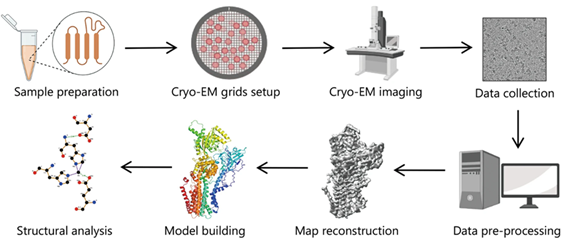 Figure 1. Typical workflow of single particle cryo-EM for structural analysis (Zhu, Kong-Fu, et al. "Applications and prospects of cryo-EM in drug discovery." Military Medical Research 10.1 (2023): 10.
Workflow of Single-Particle Cryo-EM
- Purify: Single-particle cryo-EM requires purification and structural integrity of the specimen for accurate 3D reconstructions. It necessitates maintaining the specimen in a buffer solution for biochemical activity. The molecule concentration should be high for microscopic study, while avoiding aggregation. Meanwhile, uniform molecular conformation should be ensured through optimized experimental conditions.
- Plunge freeze: The specimen is initially frozen to avoid freeze-drying within the microscope's vacuum. Rapid freezing helps prevent the disruption of specimen structure through ice crystal formation. Firstly, the specimen solution is applied to a TEM grid. Excess liquid is then blotted away briefly before the grid is submerged in liquid ethane or an ethane/propane mix. This swift wetting and cooling process forms non-crystalline or vitreous ice around the specimen. The image displays the Cryoplunge® 3 system just before blotting and specimen submersion into the liquid cryogen.
- Transfer to TEM: The frozen specimen is transitioned into a specific TEM holder that sustains it at liquid nitrogen temperature to avoid contamination. The specimen is safeguarded in a cryo-workstation during loading and encased in a cryo-shield when transferring to the TEM. The image illustrates the withdrawal of the cryo-transfer holder from the workstation, ready for TEM insertion.
- Image specimen: Specimens are vulnerable to structural damage when exposed to electrons, limiting the total dose to 10 - 30 e-/Å2 before losing high-resolution information. To mitigate this, low-dose methods are used for location and focus before image capture. Gatan's direct detector electron counting and super-resolution modes deliver high-quality, high signal-to-noise ratio images of delicate biological samples, revealing water molecules, ions, and ligand structures in the 3D particle reconstruction. Image quality can be further enhanced by utilizing the dose fractionation feature on Gatan's cameras, which captures full frames at up to 75 frames per second to later correct for specimen motion and reduce drift.
- Analyze and reconstruct: The software aids in data analysis and conversion into various formats after imaging. The data from the cameras can be incorporated into numerous third-party software tools for 3D reconstruction and visualization, such as EMAN, Frealign, Relion, etc. The displayed image is a 3D cryo-EM density of a 2.8 Å resolution 20S proteasome.
Quotation and ordering
Our customer service representatives are available 24hr a day! We thank you for considering Creative Bioarray as your Single-Particle Cryo-EM partner.
Explore Other Options
