- You are here: Home
- Services
- ISH/FISH Services
- FISH Applications
- In Situ Detection of Chemokines/Cytokines
Services
-
Cell Services
- Cell Line Authentication
- Cell Surface Marker Validation Service
-
Cell Line Testing and Assays
- Toxicology Assay
- Drug-Resistant Cell Models
- Cell Viability Assays
- Cell Proliferation Assays
- Cell Migration Assays
- Soft Agar Colony Formation Assay Service
- SRB Assay
- Cell Apoptosis Assays
- Cell Cycle Assays
- Cell Angiogenesis Assays
- DNA/RNA Extraction
- Custom Cell & Tissue Lysate Service
- Cellular Phosphorylation Assays
- Stability Testing
- Sterility Testing
- Endotoxin Detection and Removal
- Phagocytosis Assays
- Cell-Based Screening and Profiling Services
- 3D-Based Services
- Custom Cell Services
- Cell-based LNP Evaluation
-
Stem Cell Research
- iPSC Generation
- iPSC Characterization
-
iPSC Differentiation
- Neural Stem Cells Differentiation Service from iPSC
- Astrocyte Differentiation Service from iPSC
- Retinal Pigment Epithelium (RPE) Differentiation Service from iPSC
- Cardiomyocyte Differentiation Service from iPSC
- T Cell, NK Cell Differentiation Service from iPSC
- Hepatocyte Differentiation Service from iPSC
- Beta Cell Differentiation Service from iPSC
- Brain Organoid Differentiation Service from iPSC
- Cardiac Organoid Differentiation Service from iPSC
- Kidney Organoid Differentiation Service from iPSC
- GABAnergic Neuron Differentiation Service from iPSC
- Undifferentiated iPSC Detection
- iPSC Gene Editing
- iPSC Expanding Service
- MSC Services
- Stem Cell Assay Development and Screening
- Cell Immortalization
-
ISH/FISH Services
- In Situ Hybridization (ISH) & RNAscope Service
- Fluorescent In Situ Hybridization
- FISH Probe Design, Synthesis and Testing Service
-
FISH Applications
- Multicolor FISH (M-FISH) Analysis
- Chromosome Analysis of ES and iPS Cells
- RNA FISH in Plant Service
- Mouse Model and PDX Analysis (FISH)
- Cell Transplantation Analysis (FISH)
- In Situ Detection of CAR-T Cells & Oncolytic Viruses
- CAR-T/CAR-NK Target Assessment Service (ISH)
- ImmunoFISH Analysis (FISH+IHC)
- Splice Variant Analysis (FISH)
- Telomere Length Analysis (Q-FISH)
- Telomere Length Analysis (qPCR assay)
- FISH Analysis of Microorganisms
- Neoplasms FISH Analysis
- CARD-FISH for Environmental Microorganisms (FISH)
- FISH Quality Control Services
- QuantiGene Plex Assay
- Circulating Tumor Cell (CTC) FISH
- mtRNA Analysis (FISH)
- In Situ Detection of Chemokines/Cytokines
- In Situ Detection of Virus
- Transgene Mapping (FISH)
- Transgene Mapping (Locus Amplification & Sequencing)
- Stable Cell Line Genetic Stability Testing
- Genetic Stability Testing (Locus Amplification & Sequencing + ddPCR)
- Clonality Analysis Service (FISH)
- Karyotyping (G-banded) Service
- Animal Chromosome Analysis (G-banded) Service
- I-FISH Service
- AAV Biodistribution Analysis (RNA ISH)
- Molecular Karyotyping (aCGH)
- Droplet Digital PCR (ddPCR) Service
- Digital ISH Image Quantification and Statistical Analysis
- SCE (Sister Chromatid Exchange) Analysis
- Biosample Services
- Histology Services
- Exosome Research Services
- In Vitro DMPK Services
-
In Vivo DMPK Services
- Pharmacokinetic and Toxicokinetic
- PK/PD Biomarker Analysis
- Bioavailability and Bioequivalence
- Bioanalytical Package
- Metabolite Profiling and Identification
- In Vivo Toxicity Study
- Mass Balance, Excretion and Expired Air Collection
- Administration Routes and Biofluid Sampling
- Quantitative Tissue Distribution
- Target Tissue Exposure
- In Vivo Blood-Brain-Barrier Assay
- Drug Toxicity Services
In Situ Detection of Chemokines/Cytokines
Recent exciting progress in cancer immunotherapy has ushered in a new era of cancer treatment. Immunotherapy can elicit unprecedented durable response in advanced cancer patients that are much greater than conventional chemotherapy. However, such responses only occur in a relatively small fraction of patients. A positive response to immunotherapy usually relies on dynamic interactions between tumor cells and immunomodulators inside the tumor microenvironment (TME). Depending on the context of these interactions, the TME may play important roles to either dampen or enhance immune responses. Extensive studies show that some of these chemokines and cytokines participate in or represent a series of important immune and pathological processes, and the detection of these chemokines and cytokines can improve our understanding of these immunotherapeutic mechanisms. The chemokines and cytokines include the chemokine CXCL13, interleukin-6 (IL-6), interleukin 17A, interleukin 10 (IL-10), and IFNγ.
Creative Bioarray now offers the In Situ Detection of Chemokines/Cytokines service for you from the assay development, validation and final testing and analysis services. We can combine the use of immunohistochemistry and FISH for optimal detection of the chemokines/cytokines. Our team is dedicated to developing excellent services for our clients.
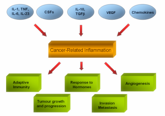
 Figure1. Role of Cytokines in cancer
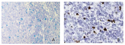
 Figure 2. A. Rare IFNG expressing infiltrating CD3+ T cell in tumor microenvironment; Hs-CD3D (Green); Hs-IFNG (Red); B. CXCL13 mRNA detection in a gastric cancer sample
Applications
- In Situ detect the distribution of chemokines/cytokines in tumor cells or tissues
- Assess the antitumor effect of chemokines/cytokines
- Confirm chemokines/cytokines expression in FFPE tissues
- Quantify chemokines/cytokines expression in FFPE tissues
Features
- Accurate-In Situ Detection Service-Custom design your probe
- Value – We focus on the quality of our service and all supported by competitive pricing
- Efficiency – We are able to provide the fastest turnaround time of any supplier in the industry
Creative Bioarray offers In Situ Detection of Chemokines/Cytokines services for your scientific research as follows:
- Probe design
- Probe synthesis
- Optimization of FISH protocols
- FISH on samples
- Imaging
- Data analysis
Quotation and ordering
Our customer service representatives are available 24hr a day! We thank you for choosing Creative Bioarray at your preferred In Situ Detection of chemokines/cytokines services.
References
- Tang H,; et al. Immunotherapy and tumor microenvironment[J]. Cancer letters, 2016, 370(1): 85-90.
- Zhang L,; et al. Lineage tracking reveals dynamic relationships of T cells in colorectal cancer[J]. Nature, 2018, 564(7735): 268.
- Tsukamoto H,; et al. Immune‐suppressive effects of interleukin‐6 on T‐cell‐mediated anti‐tumor immunity[J]. Cancer science, 2018, 109(3): 523-530.
- Thommen D S,; et al. A transcriptionally and functionally distinct PD-1+ CD8+ T cell pool with predictive potential in non-small-cell lung cancer treated with PD-1 blockade[J]. Nature medicine, 2018, 24(7): 994.
Explore Other Options
For research use only. Not for any other purpose.

