- You are here: Home
- Services
- In Vivo DMPK Services
- Quantitative Tissue Distribution
Services
-
Cell Services
- Cell Line Authentication
- Cell Surface Marker Validation Service
-
Cell Line Testing and Assays
- Toxicology Assay
- Drug-Resistant Cell Models
- Cell Viability Assays
- Cell Proliferation Assays
- Cell Migration Assays
- Soft Agar Colony Formation Assay Service
- SRB Assay
- Cell Apoptosis Assays
- Cell Cycle Assays
- Cell Angiogenesis Assays
- DNA/RNA Extraction
- Custom Cell & Tissue Lysate Service
- Cellular Phosphorylation Assays
- Stability Testing
- Sterility Testing
- Endotoxin Detection and Removal
- Phagocytosis Assays
- Cell-Based Screening and Profiling Services
- 3D-Based Services
- Custom Cell Services
- Cell-based LNP Evaluation
-
Stem Cell Research
- iPSC Generation
- iPSC Characterization
-
iPSC Differentiation
- Neural Stem Cells Differentiation Service from iPSC
- Astrocyte Differentiation Service from iPSC
- Retinal Pigment Epithelium (RPE) Differentiation Service from iPSC
- Cardiomyocyte Differentiation Service from iPSC
- T Cell, NK Cell Differentiation Service from iPSC
- Hepatocyte Differentiation Service from iPSC
- Beta Cell Differentiation Service from iPSC
- Brain Organoid Differentiation Service from iPSC
- Cardiac Organoid Differentiation Service from iPSC
- Kidney Organoid Differentiation Service from iPSC
- GABAnergic Neuron Differentiation Service from iPSC
- Undifferentiated iPSC Detection
- iPSC Gene Editing
- iPSC Expanding Service
- MSC Services
- Stem Cell Assay Development and Screening
- Cell Immortalization
-
ISH/FISH Services
- In Situ Hybridization (ISH) & RNAscope Service
- Fluorescent In Situ Hybridization
- FISH Probe Design, Synthesis and Testing Service
-
FISH Applications
- Multicolor FISH (M-FISH) Analysis
- Chromosome Analysis of ES and iPS Cells
- RNA FISH in Plant Service
- Mouse Model and PDX Analysis (FISH)
- Cell Transplantation Analysis (FISH)
- In Situ Detection of CAR-T Cells & Oncolytic Viruses
- CAR-T/CAR-NK Target Assessment Service (ISH)
- ImmunoFISH Analysis (FISH+IHC)
- Splice Variant Analysis (FISH)
- Telomere Length Analysis (Q-FISH)
- Telomere Length Analysis (qPCR assay)
- FISH Analysis of Microorganisms
- Neoplasms FISH Analysis
- CARD-FISH for Environmental Microorganisms (FISH)
- FISH Quality Control Services
- QuantiGene Plex Assay
- Circulating Tumor Cell (CTC) FISH
- mtRNA Analysis (FISH)
- In Situ Detection of Chemokines/Cytokines
- In Situ Detection of Virus
- Transgene Mapping (FISH)
- Transgene Mapping (Locus Amplification & Sequencing)
- Stable Cell Line Genetic Stability Testing
- Genetic Stability Testing (Locus Amplification & Sequencing + ddPCR)
- Clonality Analysis Service (FISH)
- Karyotyping (G-banded) Service
- Animal Chromosome Analysis (G-banded) Service
- I-FISH Service
- AAV Biodistribution Analysis (RNA ISH)
- Molecular Karyotyping (aCGH)
- Droplet Digital PCR (ddPCR) Service
- Digital ISH Image Quantification and Statistical Analysis
- SCE (Sister Chromatid Exchange) Analysis
- Biosample Services
- Histology Services
- Exosome Research Services
- In Vitro DMPK Services
-
In Vivo DMPK Services
- Pharmacokinetic and Toxicokinetic
- PK/PD Biomarker Analysis
- Bioavailability and Bioequivalence
- Bioanalytical Package
- Metabolite Profiling and Identification
- In Vivo Toxicity Study
- Mass Balance, Excretion and Expired Air Collection
- Administration Routes and Biofluid Sampling
- Quantitative Tissue Distribution
- Target Tissue Exposure
- In Vivo Blood-Brain-Barrier Assay
- Drug Toxicity Services
Quantitative Tissue Distribution
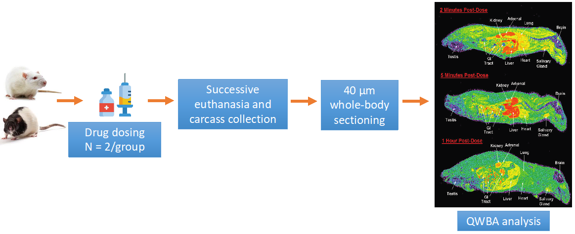
Drug concentration measurements in target and non-target tissues have been thought to be crucial for predicting drug efficacy and safety. However, extrapolation based on plasma pharmacokinetics (PK) data often fails to depict tissue exposure accurately. The quantitative tissue distribution study provides a time course of radiolabeled drug distribution and in situ concentrations in various tissues and organs via quantitative whole-body autoradiography (QWBA). In brief, animals are dosed with radiolabeled drugs, and whole-body sections are collected at successive time points for high-resolution phosphor imaging of total radioactivity.
Creative Bioarray provides quantitative tissue distribution services to help our customers visualize true tissue distribution, facilitate tissue PK analysis and dosimetry prediction before the initiation of human mass balance studies.
Animal Species
- Rodents
- Non-rodents
Mice, Rats, Guinea pigs
Dogs, Minipigs, Non-human primates
Study Design
Animals are randomly divided into different groups based on their assigned time point of euthanasia. Following is a standard study investigating the tissue distribution of a [14C]-labeled drug and related material in rats.
 Figure 1. Standard study investigation of quantitative tissue distribution assay
According to different research purposes, our experimental design can be adjusted in the following aspects, such as
- Additional animals
- Additional or custom time points
- Terminal blood plasma radioactivity quantification
- Placental transfer studies using pregnant animals
- Tumor penetration studies
- Gender effects
- Disease models
- Wet tissue radioactivity measurement in satellite animals
- Fed/food effects
- Multiple-dose studies
- Drug-drug interaction studies
- Nonradiolabeled sodium-iodide administration before 125I dosing
- Intracellular analysis via microautoradiography (MARG)
Drug dosing routes
- Default: intravenous (iv) and oral administration (po)
- Others: intraperitoneal (ip), intramuscular (im), and subcutaneous (sc) injections, iv cannulation
Dosing methods
The amount of radioactive dose is determined by the PK properties of the parent compound, typically under 100 µCi/kg. The radiolabeled drug is diluted with non radiolabeled compound to achieve the total targeted dose. While a single dose at the provided dosage is usually given to each animal, other drug dosing methods can be included to accommodate your specific needs.
- Additional dosage groups
- Repeated dosing for steady-state measurements
- Vehicle dosing
Sample collection
Sample collection typically lasts up to a week in a single-dose study but can be extended or shorten depending on the drug's half-life. A sample period covering ten plasma half-lives of the parent drug is ordinarily sufficient.
- Whole-body sections
- Section thickness normalized by quality control standards placed intro frozen blocks before sectioning
- 10-20 whole-body sections at various depths
- >40 tissues covered. Target tissue can be requested
- Blood sampling
- Tissue collection from frozen sample blocks after sectioning for QWBA
- Urine and feces
Endpoints
After at least four days of exposure, images are scanned and quantified relative to the calibration standards by image densitometry using appropriate image analysis software such as MCID. The drug concentrations derived from drug-related total radioactivity are used to generate a concentration-time profile, based on which tissue PK parameters are calculated using non-compartmental analysis. Following are general endpoints examined in a quantitative tissue distribution study and are customizable depending on your needs.
- Representative WBA images at each time point
- Concentrations of radioactivity in all significant tissues at each time point
- Tissue concentration versus time profiles
- Identified target tissues
Quotation and Ordering
As with all of our in vivo services, we ensure to keep in contact during studies. If you have any special needs or questions regarding our services, please feel free to contact us to support our experienced experts. We look forward to working with you in the future.
Reference
- FDA guidance for Industry. Safety Testing of Drug Metabolites. (March 2020)
Explore Other Options
For research use only. Not for any other purpose.

