- You are here: Home
- Disease Models
- Cardiovascular Disease Models
- Surgical Models
- Transverse Aortic Constriction (TAC) Model
Disease Models
- Oncology Models
-
Inflammation & Autoimmune Disease Models
- Rheumatoid Arthritis Models
- Glomerulonephritis Models
- Multiple Sclerosis (MS) Models
- Ocular Inflammation Models
- Sjögren's Syndrome Model
- LPS-induced Acute Lung Injury Model
- Peritonitis Models
- Passive Cutaneous Anaphylaxis Model
- Delayed-Type Hypersensitivity (DTH) Models
- Inflammatory Bowel Disease Models
- Systemic Lupus Erythematosus Animal Models
- Asthma Model
- Sepsis Model
- Psoriasis Model
- Atopic Dermatitis (AD) Model
- Scleroderma Model
- Gouty Arthritis Model
- Carrageenan-Induced Air Pouch Synovitis Model
- Carrageenan-Induced Paw Edema Model
- Experimental Autoimmune Myasthenia Gravis (EAMG) Model
-
Cardiovascular Disease Models
- Surgical Models
- Animal Models of Hypertension
- Venous Thrombosis Model
- Atherosclerosis model
- Cardiac Arrhythmia Model
- Hyperlipoidemia Model
- Doxorubicin-induced Heart Failure Model
- Isoproterenol-induced Heart Failure Model
- Arterial Thrombosis Model
- Pulmonary Arterial Hypertension (PAH) Models
- Heart Failure with Preserved Ejection Fraction (HFpEF) Model
-
Neurological Disease Models
- Alzheimer's Disease Modeling and Assays
- Seizure Models
- Parkinson's Disease Models
- Ischemic Stroke Models
- Acute Spinal Cord Injury (ASCI) Model
- Traumatic Brain Injury (TBI) Model
- Hypoxic-Ischemic Encephalopathy (HIE) Model
- Tourette Syndrome (TS) Model
- Amyotrophic Lateral Sclerosis (ALS) Model
- Huntington's Disease (HD) Model
- Intracerebral hemorrhage (ICH) Models
- Pain Models
- Metabolic Disease Models
- Liver Disease Models
- Rare Disease Models
- Respiratory Disease Models
- Digestive Disease Models
-
Urology Disease Models
- Cisplatin-induced Nephrotoxicity Model
- Unilateral Ureteral Obstruction Model
- 5/6 Nephrectomy Model
- Renal Ischemia-Reperfusion Injury (RIRI) Model
- Diabetic Nephropathy (DN) Models
- Passive Heymann Nephritis (PHN) Model
- Adenine-Induced Chronic Kidney Disease (CKD) Model
- Kidney Stone Model
- Doxorubicin-Induced Nephropathy Model
- Orthopedic Disease Models
- Ocular Disease Models
- Skin Disease Models
- Infectious Disease Models
Transverse Aortic Constriction (TAC) Model
Creative Bioarray has successfully developed a transverse aortic constriction (TAC) model, a pivotal tool in the rigorous assessment of drug candidates' efficacy against cardiac hypertrophy or chronic heart failure. This sophisticated model is meticulously designed to simulate the physiological conditions of heart diseases, enabling precise evaluation of therapeutic responses. Our team of experts employs this model to conduct comprehensive in vivo studies, providing valuable insights into the mechanisms of heart conditions and facilitating the development of more effective treatments.
The TAC model is widely recognized as a key model for studying cardiac hypertrophy and heart failure, triggered by pressure overload. Initially, TAC induces compensatory hypertrophy in the heart, sometimes accompanied by a transient improvement in cardiac contractility. However, as chronic hemodynamic stress persists, the heart's adaptive response transitions into a maladaptive state, leading to cardiac dilation and eventual heart failure. The murine TAC model is highly valued for its ability to closely replicate human cardiovascular diseases, offering insights into the critical signaling pathways involved in the progression of cardiac hypertrophy and the onset of heart failure. Compared to alternative heart failure models, such as those involving the complete blockage of the left anterior descending (LAD) coronary artery, the TAC model offers a more consistent representation of cardiac hypertrophy and a more gradual progression to heart failure, making it a preferred choice for researchers in the field.
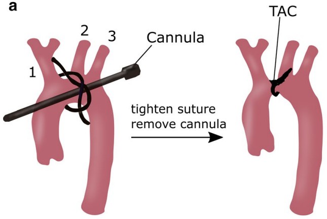
 Fig. 1 Transverse aortic constriction (TAC) in mice. (Bacmeister et al. 2019)
Our Transverse Aortic Constriction (TAC) Model
Available Animal
- Rat
- Mouse
Modeling Method
Following anesthetization, a partial thoracotomy is meticulously performed up to the second rib under the guidance of a surgical microscope. Upon successful identification of the transverse aorta, a segment of silk suture is carefully positioned between the innominate and left carotid arteries. Subsequently, two loose knots are securely tied around the transverse aorta, and a small piece of a blunted needle is positioned parallel to the transverse aorta. The first knot is swiftly tightened against the needle, followed by the second, and the needle is promptly removed to achieve a precise constriction in diameter.
 Fig. 2 Modeling method of transverse aortic constriction (TAC) model.
Endpoints
- Echocardiography
- Heart weight
- Body weight
- Histology analysis (heart): H&E staining, Masson staining
- qPCR or Western Blot
- Other customized endpoints according to your specific needs
Example Data
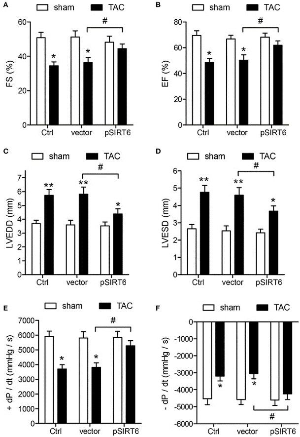
 Fig. 3 SIRT6 overexpression protects against TAC-induced impairment of cardiovascular function. Echocardiographic and hemodynamic measurements of fractional shortening (FS) (A), ejection fraction (EF) (B), left ventricular end-diastolic dimension (LVEDD) (C), left ventricular end-systolic dimension (LVESD) (D), rates of maximal rise in left ventricular pressure (+dP/dt) (E), and rate of maximal fall in left ventricular pressure (−dP/dt) (F) in control, vector and pSIRT6-injected mice at 28 days after TAC or sham surgery. (Li et al. 2017)
 Fig. 4 SIRT6 overexpression reduces infarct size and cardiac fibrosis after TAC. Myocardial tissues were collected at 28 days after surgery in control and pSIRT6-injected mice. (A) Representative images of left ventricular sections stained with Masson's trichrome at 200 × magnification. (B) Quantification of fibrotic areas. (C) Representative images of middle heart sections stained with TTC. (D) Quantitative analysis of infarct size in TAC mice from TTC stained images. (Li et al. 2017)
Quotation and Ordering
Creative Bioarray stands at the forefront of providing cutting-edge disease models tailored to meet the specific needs of our clients. Our team of in vivo pharmacology experts is dedicated to collaborating closely with you to craft a comprehensive and customized study design that aligns perfectly with your research requirements. If you are interested in our services, please feel free to contact us at any time or submit an inquiry to us directly.
References
- Li, Y., et al. Cardioprotective effects of SIRT6 in a mouse model of transverse aortic constriction-induced heart failure. Frontiers in physiology, 2017, 8: 394.
- DeAlmeida, A.C., et al. Transverse aortic constriction in mice. J Vis Exp, 2010;(38):1729.
- Bacmeister, L., et al. Inflammation and fibrosis in murine models of heart failure. Basic research in cardiology, 2019, 114: 1-35.
For research use only. Not for any other purpose.

