- You are here: Home
- Services
- Histology Services
- Transmission Electron Microscopy
- Immuno Transmission Electron Microscopy (immuno-TEM)
Services
-
Cell Services
- Cell Line Authentication
- Cell Surface Marker Validation Service
-
Cell Line Testing and Assays
- Toxicology Assay
- Drug-Resistant Cell Models
- Cell Viability Assays
- Cell Proliferation Assays
- Cell Migration Assays
- Soft Agar Colony Formation Assay Service
- SRB Assay
- Cell Apoptosis Assays
- Cell Cycle Assays
- Cell Angiogenesis Assays
- DNA/RNA Extraction
- Custom Cell & Tissue Lysate Service
- Cellular Phosphorylation Assays
- Stability Testing
- Sterility Testing
- Endotoxin Detection and Removal
- Phagocytosis Assays
- Cell-Based Screening and Profiling Services
- 3D-Based Services
- Custom Cell Services
- Cell-based LNP Evaluation
-
Stem Cell Research
- iPSC Generation
- iPSC Characterization
-
iPSC Differentiation
- Neural Stem Cells Differentiation Service from iPSC
- Astrocyte Differentiation Service from iPSC
- Retinal Pigment Epithelium (RPE) Differentiation Service from iPSC
- Cardiomyocyte Differentiation Service from iPSC
- T Cell, NK Cell Differentiation Service from iPSC
- Hepatocyte Differentiation Service from iPSC
- Beta Cell Differentiation Service from iPSC
- Brain Organoid Differentiation Service from iPSC
- Cardiac Organoid Differentiation Service from iPSC
- Kidney Organoid Differentiation Service from iPSC
- GABAnergic Neuron Differentiation Service from iPSC
- Undifferentiated iPSC Detection
- iPSC Gene Editing
- iPSC Expanding Service
- MSC Services
- Stem Cell Assay Development and Screening
- Cell Immortalization
-
ISH/FISH Services
- In Situ Hybridization (ISH) & RNAscope Service
- Fluorescent In Situ Hybridization
- FISH Probe Design, Synthesis and Testing Service
-
FISH Applications
- Multicolor FISH (M-FISH) Analysis
- Chromosome Analysis of ES and iPS Cells
- RNA FISH in Plant Service
- Mouse Model and PDX Analysis (FISH)
- Cell Transplantation Analysis (FISH)
- In Situ Detection of CAR-T Cells & Oncolytic Viruses
- CAR-T/CAR-NK Target Assessment Service (ISH)
- ImmunoFISH Analysis (FISH+IHC)
- Splice Variant Analysis (FISH)
- Telomere Length Analysis (Q-FISH)
- Telomere Length Analysis (qPCR assay)
- FISH Analysis of Microorganisms
- Neoplasms FISH Analysis
- CARD-FISH for Environmental Microorganisms (FISH)
- FISH Quality Control Services
- QuantiGene Plex Assay
- Circulating Tumor Cell (CTC) FISH
- mtRNA Analysis (FISH)
- In Situ Detection of Chemokines/Cytokines
- In Situ Detection of Virus
- Transgene Mapping (FISH)
- Transgene Mapping (Locus Amplification & Sequencing)
- Stable Cell Line Genetic Stability Testing
- Genetic Stability Testing (Locus Amplification & Sequencing + ddPCR)
- Clonality Analysis Service (FISH)
- Karyotyping (G-banded) Service
- Animal Chromosome Analysis (G-banded) Service
- I-FISH Service
- AAV Biodistribution Analysis (RNA ISH)
- Molecular Karyotyping (aCGH)
- Droplet Digital PCR (ddPCR) Service
- Digital ISH Image Quantification and Statistical Analysis
- SCE (Sister Chromatid Exchange) Analysis
- Biosample Services
- Histology Services
- Exosome Research Services
- In Vitro DMPK Services
-
In Vivo DMPK Services
- Pharmacokinetic and Toxicokinetic
- PK/PD Biomarker Analysis
- Bioavailability and Bioequivalence
- Bioanalytical Package
- Metabolite Profiling and Identification
- In Vivo Toxicity Study
- Mass Balance, Excretion and Expired Air Collection
- Administration Routes and Biofluid Sampling
- Quantitative Tissue Distribution
- Target Tissue Exposure
- In Vivo Blood-Brain-Barrier Assay
- Drug Toxicity Services
Immuno Transmission Electron Microscopy (immuno-TEM)
Immuno Transmission Electron Microscopy (immuno-TEM) is a sophisticated technique that employs immunogold labeling to detect and localize endogenous proteins within cells and tissues. By conjugating highly electron-dense gold particles, such as 10 nm gold beads, to specific primary or secondary antibodies, this method enables high-resolution and specific visualization of proteins. Immuno-TEM provides valuable insights into the distribution and quantification of proteins under various stimulation conditions, shedding light on their functional roles in cellular processes.
This technique has been instrumental in identifying the cellular and subcellular localization of proteins involved in critical biological functions, including neurotransmission and nuclear protein components, as well as in distinguishing different immune cell types. Immunolabeling can be performed on both resin sections and thawed cryo-sections, with the only requirement being that the sample retains its antigenicity throughout the processing steps to ensure accurate detection.
At Creative Bioarray, we pride ourselves on our extensive experience in Immuno Transmission Electron Microscopy (immuno-TEM). Our team of skilled professionals is dedicated to providing high-quality imaging services that enable researchers to explore the intricate details of cellular structures and protein localization.
Example of results:
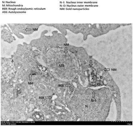
 Figure 1. Immunogold labeling for GBP1-5 in Immortalized Mouse Dendritic Cells (MutuDC1940) showed in the endoplasmic reticulum, autolysosomes, and cytoplasm. GBP1-5, 10-nm gold particles.
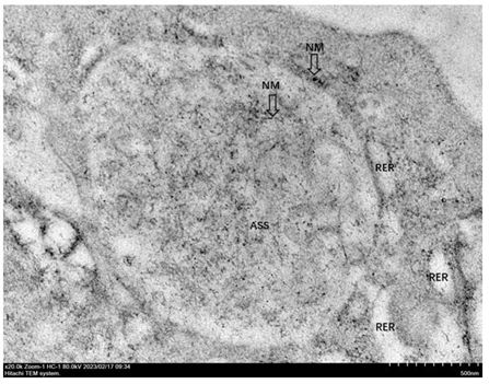
 Figure 2. Immunogold labeling for GBP1-5 in Immortalized Mouse Dendritic Cells (MutuDC1940) showed in the autolysosomes and cytoplasm. GBP1-5, 10-nm gold particles.
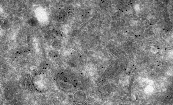
 Figure 3. Example of immunolabelled specimen. Labels can be seen as small black spots. Image courtesy Rick Webb and Rob Parton, University of Queensland.
Applications
- Identification of specific proteins and antigens in cells and tissues.
- Study of cellular localization and distribution of biomolecules.
- Research in cell biology, pathology, and immunology.
- Analysis of disease mechanisms at the ultrastructural level.
Advantages
- High-resolution imaging for detailed structural analysis.
- Specificity in targeting proteins using antibodies.
- Ability to visualize complex cellular interactions.
- Comprehensive data for research and diagnostic purposes.
Service Process
- Consultation: Discuss your research needs and sample requirements with our experts.
- Sample Preparation: We assist in preparing your samples for optimal results.
- Immunolabeling: Specific antibodies are applied to target the proteins of interest.
- Negative Staining: Enhance contrast for better visualization under the electron microscope.
- TEM Imaging: High-resolution imaging of your samples to capture detailed structures.
- Data Analysis: Comprehensive analysis and reporting of the findings.
Quotation and ordering
Our customer service representatives are available 24hr a day! We thank you for choosing Creative Bioarray at your preferred Immuno Transmission Electron Microscopy (immuno-TEM) Services.
FAQs
1. What is Immuno-TEM?
Immuno Transmission Electron Microscopy (immuno-TEM) is a technique used to localize antigens in cells and tissues by employing antigen-specific primary antibodies in combination with electron-dense markers, typically colloidal gold. This method allows for high-resolution visualization of proteins and other biomolecules within cellular structures.
2. How does the immuno-TEM process work?
The immuno-TEM process generally involves a two-step detection method. Initially, primary antibodies bind to the target antigens. Subsequently, gold-conjugated secondary antibodies, or proteins such as Protein A or Protein G, are used to enhance the signal, allowing for the visualization of the antigen-antibody complexes under an electron microscope.
3. What types of gold reagents are available for immuno-TEM?
We offer a wide range of gold reagents for immuno-TEM, including conventional gold-conjugated antibodies with particle sizes of 6, 10, 15, or 25 nm, as well as ultra-small immunogold conjugates prepared with subnanometer gold particles. The ultra-small reagents are suitable for both immuno-electron microscopy and immuno-light microscopy, minimizing the impact on the adsorbed antibodies.
4. How can I order gold-labeled antibodies or reagents?
You can easily order your gold-labeled antibodies or other reagents directly from our webshop. We ensure prompt delivery within three working days. If you require a custom conjugate, you can also request our custom labeling service.
5. How can I contact you for more information?
For any questions about our products or services, feel free to contact us by calling 1-631-626-9181(USA) or 44-208-123-7131(Europe) or sending an email to info@creative-bioarray.com. We are here to assist you!
Explore Other Options
For research use only. Not for any other purpose.

