Arterial Thrombosis Model
Creative Bioarray stands as a premier and dependable industry pioneer in the realm of developing animal models for various diseases. Specifically, we have established a robust and reproducible model of ferric chloride (FeCl3)-induced arterial thrombosis, which serves as a pivotal tool in preclinical research for screening potential drug candidates and conducting in vivo efficacy studies. Our commitment to excellence in this domain ensures the highest level of accuracy and reproducibility, thus enhancing the reliability of our research outcomes.
Arterial thrombosis, or the formation of blood clots in arteries, is a frequent complication arising from numerous chronic inflammatory systemic diseases, such as atherosclerosis, diabetes, obesity, cancer, and chronic autoimmune rheumatologic disorders. The FeCl3-induced arterial thrombosis model is a universally recognized and extensively employed experimental paradigm for studying thrombosis. Upon application to the exterior surface of blood vessels, FeCl3 initiates oxidative damage to vascular cells, resulting in the loss of endothelial cell-mediated protection against circulating platelets and components of the coagulation cascade. Notably, the FeCl3 model exhibits simplicity and sensitivity, making it responsive to both anticoagulants and anti-platelet agents, thereby rendering it a valuable tool in drug discovery and research.
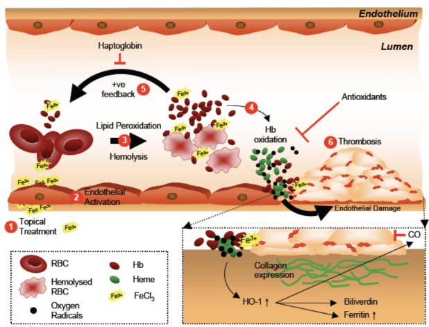 Fig. 1 Schematic illustration of the FeCl3 -induced hemolysis/Hb oxidation cycle that induces further red cell damage, vascular injury, and thrombosis. (Woollard et al. 2009)
Our Arterial Thrombosis Model
- Available Animal
Rat
- Modeling Method
After anesthetization, the abdominal artery or carotid artery is dissected free from surrounding tissue. A piece of filter paper with FeCl3 solution is placed on the surface of the artery.
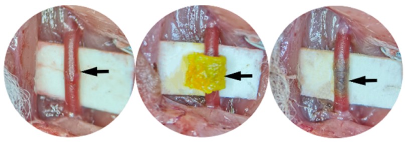 Fig. 2 Schematic diagram of arterial thrombosis induced by FeCl3. (Lin et al. 2022)
- Endpoints
- Weight of thrombus
- Length of thrombus
- Oxidative damager analysis
- Histology analysis
- qPCR or Western blot
- Other customized endpoints
Example Data
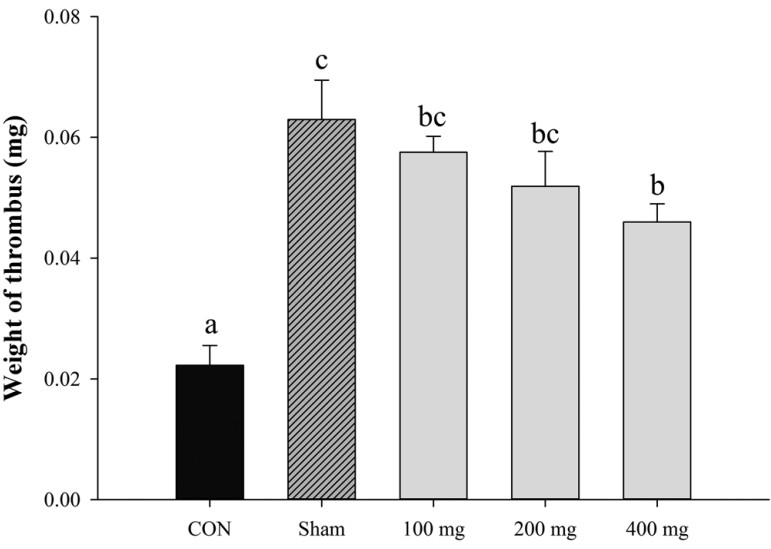 Fig. 3 Effect of subcritical water extraction (SWE) of Artemisia princeps Pampanini in FeCl3-induced carotid artery thrombosis model: measurement of thrombus formation. (Kim et al. 2019)
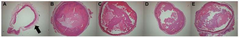 Fig. 4 H&E staining of an FeCl3-induced thrombus from the (A) Normal control, (B) Sham control, (C) subcritical water extraction (SWE) 100 mg/kg BW, (D) SWE 200 mg/kg BW, and (E) SWE 400 mg/kg BW. (Kim et al. 2019)
Furthermore, we also provide other thrombosis models that maybe you are interested in:
Quotation and Ordering
Creative Bioarray is pleased to offer our state-of-the-art technology and extensive expertise in rodent disease models to support our clients' research and project development endeavors. If you are interested in our services, please feel free to contact us at any time or submit an inquiry to us directly.
References
- Kim, K.J., et al. Antithrombotic Effect of Artemisia princeps Pampanini Extracts in Vitro and in FeCl3-Induced Thrombosis Rats. J Food Sci. 2019; 84(10):3037-3044.
- Lin, X., et al. Establishment of a Modified and Standardized Ferric Chloride-Induced Rat Carotid Artery Thrombosis Model, ACS Omega. 2022;7(10):8919-8927.
- Woollard, K.J., et al. Erythrocyte hemolysis and hemoglobin oxidation promote ferric chloride-induced vascular injury. J Biol Chem, 2009;284(19):13110-13118.