- You are here: Home
- Applications
- Skin
- Cosmetics
- Photo-aging & UV-Protection
Applications
-
Cell Services
- Cell Line Authentication
- Cell Surface Marker Validation Service
-
Cell Line Testing and Assays
- Toxicology Assay
- Drug-Resistant Cell Models
- Cell Viability Assays
- Cell Proliferation Assays
- Cell Migration Assays
- Soft Agar Colony Formation Assay Service
- SRB Assay
- Cell Apoptosis Assays
- Cell Cycle Assays
- Cell Angiogenesis Assays
- DNA/RNA Extraction
- Custom Cell & Tissue Lysate Service
- Cellular Phosphorylation Assays
- Stability Testing
- Sterility Testing
- Endotoxin Detection and Removal
- Phagocytosis Assays
- Cell-Based Screening and Profiling Services
- 3D-Based Services
- Custom Cell Services
- Cell-based LNP Evaluation
-
Stem Cell Research
- iPSC Generation
- iPSC Characterization
-
iPSC Differentiation
- Neural Stem Cells Differentiation Service from iPSC
- Astrocyte Differentiation Service from iPSC
- Retinal Pigment Epithelium (RPE) Differentiation Service from iPSC
- Cardiomyocyte Differentiation Service from iPSC
- T Cell, NK Cell Differentiation Service from iPSC
- Hepatocyte Differentiation Service from iPSC
- Beta Cell Differentiation Service from iPSC
- Brain Organoid Differentiation Service from iPSC
- Cardiac Organoid Differentiation Service from iPSC
- Kidney Organoid Differentiation Service from iPSC
- GABAnergic Neuron Differentiation Service from iPSC
- Undifferentiated iPSC Detection
- iPSC Gene Editing
- iPSC Expanding Service
- MSC Services
- Stem Cell Assay Development and Screening
- Cell Immortalization
-
ISH/FISH Services
- In Situ Hybridization (ISH) & RNAscope Service
- Fluorescent In Situ Hybridization
- FISH Probe Design, Synthesis and Testing Service
-
FISH Applications
- Multicolor FISH (M-FISH) Analysis
- Chromosome Analysis of ES and iPS Cells
- RNA FISH in Plant Service
- Mouse Model and PDX Analysis (FISH)
- Cell Transplantation Analysis (FISH)
- In Situ Detection of CAR-T Cells & Oncolytic Viruses
- CAR-T/CAR-NK Target Assessment Service (ISH)
- ImmunoFISH Analysis (FISH+IHC)
- Splice Variant Analysis (FISH)
- Telomere Length Analysis (Q-FISH)
- Telomere Length Analysis (qPCR assay)
- FISH Analysis of Microorganisms
- Neoplasms FISH Analysis
- CARD-FISH for Environmental Microorganisms (FISH)
- FISH Quality Control Services
- QuantiGene Plex Assay
- Circulating Tumor Cell (CTC) FISH
- mtRNA Analysis (FISH)
- In Situ Detection of Chemokines/Cytokines
- In Situ Detection of Virus
- Transgene Mapping (FISH)
- Transgene Mapping (Locus Amplification & Sequencing)
- Stable Cell Line Genetic Stability Testing
- Genetic Stability Testing (Locus Amplification & Sequencing + ddPCR)
- Clonality Analysis Service (FISH)
- Karyotyping (G-banded) Service
- Animal Chromosome Analysis (G-banded) Service
- I-FISH Service
- AAV Biodistribution Analysis (RNA ISH)
- Molecular Karyotyping (aCGH)
- Droplet Digital PCR (ddPCR) Service
- Digital ISH Image Quantification and Statistical Analysis
- SCE (Sister Chromatid Exchange) Analysis
- Biosample Services
- Histology Services
- Exosome Research Services
- In Vitro DMPK Services
-
In Vivo DMPK Services
- Pharmacokinetic and Toxicokinetic
- PK/PD Biomarker Analysis
- Bioavailability and Bioequivalence
- Bioanalytical Package
- Metabolite Profiling and Identification
- In Vivo Toxicity Study
- Mass Balance, Excretion and Expired Air Collection
- Administration Routes and Biofluid Sampling
- Quantitative Tissue Distribution
- Target Tissue Exposure
- In Vivo Blood-Brain-Barrier Assay
- Drug Toxicity Services
Photo-aging & UV-Protection


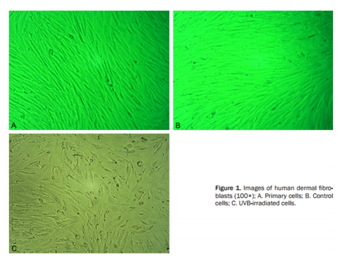
Exposure to solar UV radiation (UVA, thermal component, UVB) is the main environmental factor that causes premature aging of the skin (photo-aging). Human skin aging resulting from UV irradiation is a cumulative process that occurs based on the degree of sun exposure and the level of skin pigment. UV irradiation induces matrix metalloproteinases (MMPs) responsible for alterations in the collagenous extracellular matrix of connective tissue, resulting in impaired integrity. On a molecular level, UV radiation from the sun attacks keratinocytes and fibroblasts, resulting in the activation of cell surface receptors, which initiate signal transduction cascades. This in turn leads to a variety of molecular changes, which causes a breakdown of collagen in the extracellular matrix and a shutdown of new collagen synthesis.
In order to identify most efficient applications of the new ingredients of UV-Protection, the need for in vitro testing in cosmetic or dermatological industries is greatly growing nowadays, because of below aspects: firstly, regulatory affairs that include consumer safety and strict ban of animal use in testing. Secondly, there is a big advantage regarding research and development because of faster screening and access to more informative tests. Finally, this leads to on the evidence based communication with consumers.
Your Needs
- You wish to screen ingredients, and characterize the efficacy or toxicity or safety of any products or chemicals of UV-protection?
- You'd like to find a customized in vitro testing service of photo-aging or UV-protection?
Our Capability
Creative Bioarray provides in vitro 3D skin tissue models and evaluation assays performed for cosmetic or dermatological efficacy and claims support of photo-aging & UV protection.
In vitro 3D skin models available
- Normal human epidermal keratinocytes (NHEK)
- Reconstructed human epidermis (RHE)
- Ex vivo skin explants
- Photo-ageing model
Assays available
- Phototoxicity Assay (OECD Test Guideline 432)
- Evaluating effects of different formulations, ingredients by using 3D skin models
- Other customized assays by using 3D skin models
Endpoints
- Inflammatory mediator release (IL-8, PGE2, IL-6, TNF-alpha, etc.)
- Free radical production
- Stress response
- Metalloproteinase expression or production
- Dermal-epidermal junction markers: Integrin β4, Laminin 5
- Apoptosis
- Cell viability or metabolism
Techniques
- qPCR, qPCRarray, RT-PCR
- Epidermal separation
- Immunofluorescence
- ELISA
- RNA extraction
- Protein extraction
- Macroscopy
Assay Examples
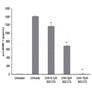
 Fig 1. Cosmetic ingredient inhibits UVA-induced MMP-1 release
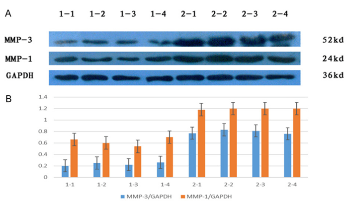
 Fig 2. Comparison of relative expression of MMP-1 and MMP-3 in UVB group and control group.
Related Products and Services
Choose our models to perform screening assays in house, or choose our assays and services directly!!
- Normal human epidermal keratinocytes(NHEK)
- Reconstructed human epidermis (RHE)
- Ex vivo skin explants
- Compound screening service
Our customer service representatives are available 24hr a day! We thank you for choosing Creative Bioarray services!
In vitro Skin Models:
Explore Other Options
For research use only. Not for any other purpose.

