- You are here: Home
- Applications
- Skin
- In vitro Skin Models
- Ex vivo Skin Explants
Applications
-
Cell Services
- Cell Line Authentication
- Cell Surface Marker Validation Service
-
Cell Line Testing and Assays
- Toxicology Assay
- Drug-Resistant Cell Models
- Cell Viability Assays
- Cell Proliferation Assays
- Cell Migration Assays
- Soft Agar Colony Formation Assay Service
- SRB Assay
- Cell Apoptosis Assays
- Cell Cycle Assays
- Cell Angiogenesis Assays
- DNA/RNA Extraction
- Custom Cell & Tissue Lysate Service
- Cellular Phosphorylation Assays
- Stability Testing
- Sterility Testing
- Endotoxin Detection and Removal
- Phagocytosis Assays
- Cell-Based Screening and Profiling Services
- 3D-Based Services
- Custom Cell Services
- Cell-based LNP Evaluation
-
Stem Cell Research
- iPSC Generation
- iPSC Characterization
-
iPSC Differentiation
- Neural Stem Cells Differentiation Service from iPSC
- Astrocyte Differentiation Service from iPSC
- Retinal Pigment Epithelium (RPE) Differentiation Service from iPSC
- Cardiomyocyte Differentiation Service from iPSC
- T Cell, NK Cell Differentiation Service from iPSC
- Hepatocyte Differentiation Service from iPSC
- Beta Cell Differentiation Service from iPSC
- Brain Organoid Differentiation Service from iPSC
- Cardiac Organoid Differentiation Service from iPSC
- Kidney Organoid Differentiation Service from iPSC
- GABAnergic Neuron Differentiation Service from iPSC
- Undifferentiated iPSC Detection
- iPSC Gene Editing
- iPSC Expanding Service
- MSC Services
- Stem Cell Assay Development and Screening
- Cell Immortalization
-
ISH/FISH Services
- In Situ Hybridization (ISH) & RNAscope Service
- Fluorescent In Situ Hybridization
- FISH Probe Design, Synthesis and Testing Service
-
FISH Applications
- Multicolor FISH (M-FISH) Analysis
- Chromosome Analysis of ES and iPS Cells
- RNA FISH in Plant Service
- Mouse Model and PDX Analysis (FISH)
- Cell Transplantation Analysis (FISH)
- In Situ Detection of CAR-T Cells & Oncolytic Viruses
- CAR-T/CAR-NK Target Assessment Service (ISH)
- ImmunoFISH Analysis (FISH+IHC)
- Splice Variant Analysis (FISH)
- Telomere Length Analysis (Q-FISH)
- Telomere Length Analysis (qPCR assay)
- FISH Analysis of Microorganisms
- Neoplasms FISH Analysis
- CARD-FISH for Environmental Microorganisms (FISH)
- FISH Quality Control Services
- QuantiGene Plex Assay
- Circulating Tumor Cell (CTC) FISH
- mtRNA Analysis (FISH)
- In Situ Detection of Chemokines/Cytokines
- In Situ Detection of Virus
- Transgene Mapping (FISH)
- Transgene Mapping (Locus Amplification & Sequencing)
- Stable Cell Line Genetic Stability Testing
- Genetic Stability Testing (Locus Amplification & Sequencing + ddPCR)
- Clonality Analysis Service (FISH)
- Karyotyping (G-banded) Service
- Animal Chromosome Analysis (G-banded) Service
- AAV Biodistribution Analysis (RNA ISH)
- Molecular Karyotyping (aCGH)
- Droplet Digital PCR (ddPCR) Service
- Digital ISH Image Quantification and Statistical Analysis
- SCE (Sister Chromatid Exchange) Analysis
- Biosample Services
- Histology Services
- Exosome Research Services
- In Vitro DMPK Services
-
In Vivo DMPK Services
- Pharmacokinetic and Toxicokinetic
- PK/PD Biomarker Analysis
- Bioavailability and Bioequivalence
- Bioanalytical Package
- Metabolite Profiling and Identification
- In Vivo Toxicity Study
- Mass Balance, Excretion and Expired Air Collection
- Administration Routes and Biofluid Sampling
- Quantitative Tissue Distribution
- Target Tissue Exposure
- In Vivo Blood-Brain-Barrier Assay
- Drug Toxicity Services
Ex vivo Skin Explants
Animal models have been proven to be not perfect for dermocosmetics testing, since animal models do not accurately represent the structure of human skin. In vitro, ex vivo techniques can aid skin research which can be adapted to study a range of conditions in order to more accurately study human skin diseases and aid drug development.
Compared with in vitro skin models, ex vivo models can be more accurate on representing the human skin in some case, such as the effect of the extracellular matrix (ECM) in terms of structure, cell signaling mechanisms and the processes of metabolism, the incorporation of skin appendages such as hair follicles and sweat glands, and the effects of dermal absorption. Ex vivo human skin is more appropriate for certain types of research, use of whole skin biopsies in culture allows the effect of individual ingredients and formulations, transdermal delivery, topical penetration and percutaneous absorption to be tested in an environment more closely mimicking normal skin.
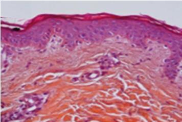
Fig.1 Histological examination of H&E-stained skin cryosections showing the morphology of the stratum corneum, epidermis, and dermis.
Creative Bioarray provides highly predictive and cost-effective ex vivo human skin model and various ingredients and formulation screening services in laboratory conditions prior to in vivo clinical evaluations for your cosmetics and dermatology products.
Creative Bioarray ex vivo human skin model advantages
- The skin model closest to clinical testing
- Skin excised from different body areas
- Inter-individual variability can be tested
Specific Markers
- Filaggrin
- Involucrin
- Loricrin
- Corneodesmosin
- Procollagne 1
- Tropoelastin
- Decorin
- Perlecan
- Keratin 10
- Keratin 5
- Presence of different epidermal classes of lipids comprising ceramides
- Collagen
- Laminin V
- Alpha6Beta4-integrin
- BP antigen
- Ki67
- Others
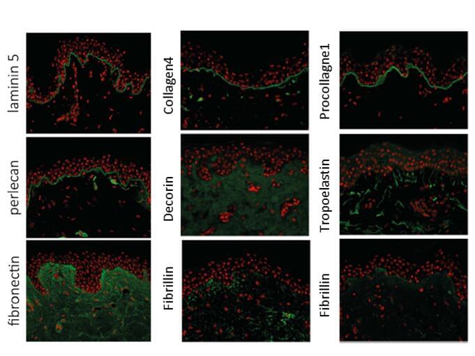
Fig.3 Immuno-labeling of different markers in ex vivo skin explant
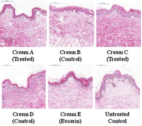
Fig.2 Immunohistological analysis of tissue morphology in
an organ culture model using hematoxylin and eosin (H and E) staining
Applications
Creative Bioarray ex vivo skin explants allows for the evaluation of topically applied compounds, chemicals, cosmetic/personal care product ingredients and final formulations for the following supporting claims:
- Anti Aging
- Pigmentation
- Stress / Inflammation
- UV Protection
Our ex vivo skin explants can be applicable to various testing including Efficacy testing, Topical application, Dermal studies, Epidermal studies, Percutaneous absorption, Metabolism, Long term studies, Repeated dose assay, Langerhans cells study, Skin resident T cells study, Immune responses, Melanogenesis, and Melanocyte study Permeability.
Quote and ordering
Our customer service representatives are available 24hr a day!
Related models
Reconstructed Human Epidermis (RHE)
In vitro Full Thickness Skin Model
Neurodermatology Skin Model
Pigmented Epidermis Model
Psoriasis Skin Model
Explore Other Options
For research use only. Not for any other purpose.