- You are here: Home
- Disease Models
- Oncology Models
- Orthotopic Models
Disease Models
- Oncology Models
-
Inflammation & Autoimmune Disease Models
- Rheumatoid Arthritis Models
- Glomerulonephritis Models
- Multiple Sclerosis (MS) Models
- Ocular Inflammation Models
- Sjögren's Syndrome Model
- LPS-induced Acute Lung Injury Model
- Peritonitis Models
- Passive Cutaneous Anaphylaxis Model
- Delayed-Type Hypersensitivity (DTH) Models
- Inflammatory Bowel Disease Models
- Systemic Lupus Erythematosus Animal Models
- Asthma Model
- Sepsis Model
- Psoriasis Model
- Atopic Dermatitis (AD) Model
- Scleroderma Model
- Gouty Arthritis Model
- Carrageenan-Induced Air Pouch Synovitis Model
- Carrageenan-Induced Paw Edema Model
- Experimental Autoimmune Myasthenia Gravis (EAMG) Model
-
Cardiovascular Disease Models
- Surgical Models
- Animal Models of Hypertension
- Venous Thrombosis Model
- Atherosclerosis model
- Cardiac Arrhythmia Model
- Hyperlipoidemia Model
- Doxorubicin-induced Heart Failure Model
- Isoproterenol-induced Heart Failure Model
- Arterial Thrombosis Model
- Pulmonary Arterial Hypertension (PAH) Models
- Heart Failure with Preserved Ejection Fraction (HFpEF) Model
-
Neurological Disease Models
- Alzheimer's Disease Modeling and Assays
- Seizure Models
- Parkinson's Disease Models
- Ischemic Stroke Models
- Acute Spinal Cord Injury (ASCI) Model
- Traumatic Brain Injury (TBI) Model
- Hypoxic-Ischemic Encephalopathy (HIE) Model
- Tourette Syndrome (TS) Model
- Amyotrophic Lateral Sclerosis (ALS) Model
- Huntington's Disease (HD) Model
- Intracerebral hemorrhage (ICH) Models
- Pain Models
- Metabolic Disease Models
- Liver Disease Models
- Rare Disease Models
- Respiratory Disease Models
- Digestive Disease Models
-
Urology Disease Models
- Cisplatin-induced Nephrotoxicity Model
- Unilateral Ureteral Obstruction Model
- 5/6 Nephrectomy Model
- Renal Ischemia-Reperfusion Injury (RIRI) Model
- Diabetic Nephropathy (DN) Models
- Passive Heymann Nephritis (PHN) Model
- Adenine-Induced Chronic Kidney Disease (CKD) Model
- Kidney Stone Model
- Doxorubicin-Induced Nephropathy Model
- Orthopedic Disease Models
- Ocular Disease Models
- Skin Disease Models
- Infectious Disease Models
Orthotopic Models
Creative Bioarray provides an extensive portfolio of orthotopic models that encompass a wide spectrum of tumor histotypes, ensuring that researchers have access to the most physiologically relevant tumor environments for the rigorous testing and validation of their therapeutic candidates. Our commitment to offering a diverse array of models is designed to closely replicate the intricacies of human disease, thereby enhancing the predictive value and translational potential of preclinical.
Introduction of Orthotopic Models
Orthotopic models are advanced preclinical tools that involve the implantation of tumor cells directly into the organ of origin, allowing for a more accurate representation of the tumor microenvironment and interactions with surrounding stromal and immune cells. By utilizing Luciferase gene-transfected cells, these models enable non-invasive bioluminescence imaging to monitor tumor growth, distribution, and metastasis in real-time. Orthotopic models closely mimic the human disease process, providing valuable insights into therapeutic efficacy by enabling the assessment of primary tumor growth, metastatic behavior, and response to treatments in a clinically relevant context. Their high predictive therapeutic value makes them essential for meaningful drug testing and evaluation.
Key Advantages of Orthotopic Models
- Interaction with the Immune System
Orthotopic models serve as a critical platform for investigating the dynamic interactions between tumors and the immune system, offering deeper insights into how these interactions influence cancer progression and treatment outcomes.
- Enhanced Clinical Relevance
By examining tumors within their original context, the orthotopic models ensure that research outcomes are highly relevant to clinical settings. It serves as a crucial link between preclinical research and human clinical trials, thereby expediting the discovery and validation of effective cancer treatments.
- Natural Tumor Microenvironment
Orthotopic models closely simulate the natural conditions of a tumor's growth environment, including specific tissue factors, blood supply, and interactions with adjacent cells. This setup allows for the observation of tumor behavior in a setting that closely mirrors actual clinical conditions.
Orthotopic Models in Creative Bioarray
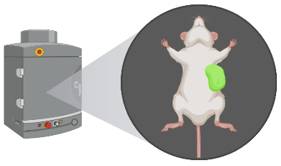
Creative Bioarray has honed its expertise over the past 10 years in orthotopic tumor implantation, a procedure that often necessitates surgical precision. Our team of seasoned veterinary surgeons is adept in a variety of surgical techniques, enabling us to meticulously implant tumors into a diverse array of organs. This proficiency has allowed us to develop a comprehensive suite of orthotopic models that accurately replicate the complexities of tumor growth within specific organ environments.
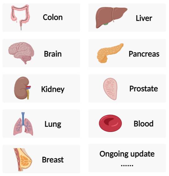 Fig. 1 Creative Bioarray's orthotopic models.
We possess a multifaceted approach to assessing disease progression, leveraging an array of sophisticated techniques. These include monitoring clinical signs, conducting survival studies, and utilizing our advanced imaging platform. Our capabilities encompass:
- Imaging: Real-time in-life fluorescence and bioluminescence imaging, a non-invasive technique, enables the monitoring of tumor growth and metastasis in real-time, offering insights into treatment efficacy and serving as a method for identifying biomarkers for response or resistance.
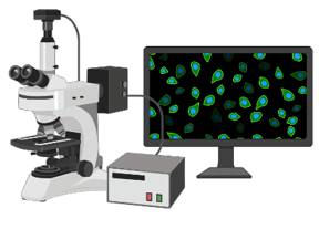
- Histology and Immunohistochemistry: Histological techniques can be used to analyze tumor tissues within an orthotopic mouse model. This allows for the examination of changes in tissue architecture, cell morphology, and protein expression, which provide valuable insights into the biological effects of treatments.
Example Data
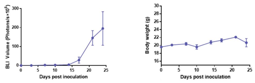 Fig. 2 Bioluminescence imaging (BLI) volume and body weight monitoring in the U87MG-Luc2 brain orthotopic tumor model.
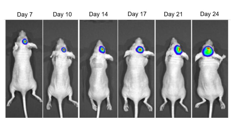 Fig. 3 In vivo imaging of the U87MG-Luc2 brain orthotopic tumor model.
Quotation and Ordering
Creative Bioarray is committed to offering a comprehensive suite of well-established models tailored for cutting-edge tumor research, ensuring that our clients have access to the most advanced tools and platforms to facilitate their scientific discoveries and therapeutic advancements. If you are interested in our services, please feel free to contact us at any time or submit an inquiry to us directly.
For research use only. Not for any other purpose.
