- You are here: Home
- Services
- In Vitro DMPK Services
- In Vitro Permeability and Transporters
- Parallel Artificial Membrane Permeability Assay (PAMPA)
Services
-
Cell Services
- Cell Line Authentication
- Cell Surface Marker Validation Service
-
Cell Line Testing and Assays
- Toxicology Assay
- Drug-Resistant Cell Models
- Cell Viability Assays
- Cell Proliferation Assays
- Cell Migration Assays
- Soft Agar Colony Formation Assay Service
- SRB Assay
- Cell Apoptosis Assays
- Cell Cycle Assays
- Cell Angiogenesis Assays
- DNA/RNA Extraction
- Custom Cell & Tissue Lysate Service
- Cellular Phosphorylation Assays
- Stability Testing
- Sterility Testing
- Endotoxin Detection and Removal
- Phagocytosis Assays
- Cell-Based Screening and Profiling Services
- 3D-Based Services
- Custom Cell Services
- Cell-based LNP Evaluation
-
Stem Cell Research
- iPSC Generation
- iPSC Characterization
-
iPSC Differentiation
- Neural Stem Cells Differentiation Service from iPSC
- Astrocyte Differentiation Service from iPSC
- Retinal Pigment Epithelium (RPE) Differentiation Service from iPSC
- Cardiomyocyte Differentiation Service from iPSC
- T Cell, NK Cell Differentiation Service from iPSC
- Hepatocyte Differentiation Service from iPSC
- Beta Cell Differentiation Service from iPSC
- Brain Organoid Differentiation Service from iPSC
- Cardiac Organoid Differentiation Service from iPSC
- Kidney Organoid Differentiation Service from iPSC
- GABAnergic Neuron Differentiation Service from iPSC
- Undifferentiated iPSC Detection
- iPSC Gene Editing
- iPSC Expanding Service
- MSC Services
- Stem Cell Assay Development and Screening
- Cell Immortalization
-
ISH/FISH Services
- In Situ Hybridization (ISH) & RNAscope Service
- Fluorescent In Situ Hybridization
- FISH Probe Design, Synthesis and Testing Service
-
FISH Applications
- Multicolor FISH (M-FISH) Analysis
- Chromosome Analysis of ES and iPS Cells
- RNA FISH in Plant Service
- Mouse Model and PDX Analysis (FISH)
- Cell Transplantation Analysis (FISH)
- In Situ Detection of CAR-T Cells & Oncolytic Viruses
- CAR-T/CAR-NK Target Assessment Service (ISH)
- ImmunoFISH Analysis (FISH+IHC)
- Splice Variant Analysis (FISH)
- Telomere Length Analysis (Q-FISH)
- Telomere Length Analysis (qPCR assay)
- FISH Analysis of Microorganisms
- Neoplasms FISH Analysis
- CARD-FISH for Environmental Microorganisms (FISH)
- FISH Quality Control Services
- QuantiGene Plex Assay
- Circulating Tumor Cell (CTC) FISH
- mtRNA Analysis (FISH)
- In Situ Detection of Chemokines/Cytokines
- In Situ Detection of Virus
- Transgene Mapping (FISH)
- Transgene Mapping (Locus Amplification & Sequencing)
- Stable Cell Line Genetic Stability Testing
- Genetic Stability Testing (Locus Amplification & Sequencing + ddPCR)
- Clonality Analysis Service (FISH)
- Karyotyping (G-banded) Service
- Animal Chromosome Analysis (G-banded) Service
- I-FISH Service
- AAV Biodistribution Analysis (RNA ISH)
- Molecular Karyotyping (aCGH)
- Droplet Digital PCR (ddPCR) Service
- Digital ISH Image Quantification and Statistical Analysis
- SCE (Sister Chromatid Exchange) Analysis
- Biosample Services
- Histology Services
- Exosome Research Services
- In Vitro DMPK Services
-
In Vivo DMPK Services
- Pharmacokinetic and Toxicokinetic
- PK/PD Biomarker Analysis
- Bioavailability and Bioequivalence
- Bioanalytical Package
- Metabolite Profiling and Identification
- In Vivo Toxicity Study
- Mass Balance, Excretion and Expired Air Collection
- Administration Routes and Biofluid Sampling
- Quantitative Tissue Distribution
- Target Tissue Exposure
- In Vivo Blood-Brain-Barrier Assay
- Drug Toxicity Services
Parallel Artificial Membrane Permeability Assay (PAMPA)
Creative Bioarray is a reliable Parallel Artificial Membrane Permeability Assay (PAMPA) provider. Over the past years, Creative Bioarray has developed expertise in PAMPA. By working with a variety of customers, we can perfectly meet your project requirements and budgets.
PAMPA Introduction
- Diffusion and absorption of drugs
Drug permeability is one of the most important factors to be considered for predicting oral drug bioavailability. Permeation mechanisms through biological barriers include active transport, passive diffusion, paracellular, and efflux. The body absorbs nearly 80~95% of the recorded commercial drugs through passive diffusion.
For permeability screening, the Caco-2 cell monolayer permeation method has been widely and successfully used in drug discovery and early development. With the need for reduced cost, increased predictability, and higher throughput in drug discovery, other methods are needed to assist Caco-2 assay.
- Why PAMPA?
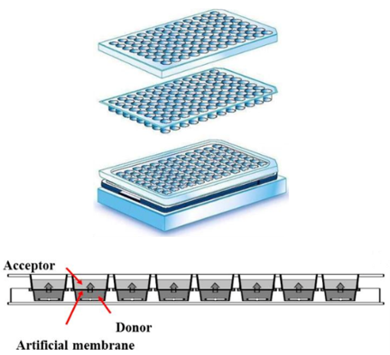
PAMPA uses two aqueous buffer solution holes separated by an artificial membrane. The artificial membrane is composed of a lipid-oil-lipid sandwich structure in an organic diluent supported by a porous filter plate matrix. And then, the test compound diluted in the buffer is placed in the donor well. The compound enters the artificial membrane from the donor pore, entering the acceptor pore by passive diffusion. The compound's effective permeability (Pe) is utilized to determine the rate of permeation. Compared with cell-monolayer methods, the time required for the experiment is greatly reduced. Only passive diffusion is tested, and there is no metabolism, no transporter proteins, so there is no need to worry about saturation.
Key features
- Improvement of correlation with human absorption and Caco-2
- Lower mass retention
- High stability and reproducibility
- Compatible with buffers containing organic solvents
- High throughput
- No metabolism
- Rapid quantitation
- Low cost
PAMPA Methods
- GIT-PAMPA
- BBB-PAMPA
- Skin-PAMPA
Brief Protocol
The PAMPA permeability test is based on the passive diffusion of the target compound through the artificial membrane. PAMPA synthetic membrane has a lipid-oil-lipid sandwich structure built into the pores of the porous filter. The middle oil layer maintains a strong and stable PAMPA membrane, ultra-thin to minimize compound retention and interference with compound penetration (Avdeef, 2005; Kerns et al., 2004).
 Figure 1. The schematic diagram of the PAMPA model
Study example
- Preparation of PAMPA Assay
Compound donor solutions were added to each well of the donor plate, whose PVDF membrane was precoated with 5 µL of 1% lecithin/dodecane mixture. 300 µL of PBS was added to each well of the PTFE acceptor plate. The donor plate and acceptor plate were combined and incubated for 4h.
 Figure 2. PAMPA Model.
- Method Validation
The apparent permeability and recovery of the test compounds were determined in duplicate. Compounds were quantified by LC-MS/MS analysis based on the peak area.
| PAMPA Test Results | |||||
| Compound | Test Conc. (µM) | Incubation Time (hour) | Mean Pe (nm/s) | Mean %Recovery | Permeability |
| Atenolol | 10 | 4 | <0.184 | <107.1 | Low |
| Propranolol | 10 | 4 | 59.9 | 69.0 | High |
Note: [drug]equilibrium=([drug]donor×VD+[drug]acceptor×VA)/(VD+VA) [drug]equilibrium=([drug]donor×VD+[drug]acceptor×VA)/(VD+VA)VD = 0.15 mL; VA = 0.30 mL; Area = 0.28 cm2; time = 14400 s. [drug]acceptor = (Aa/Ai×DF)acceptor; [drug]donor= (Aa/Ai*DF)donor; Aa/Ai: Peak area ratio of analyte and internal standard; DF: Dilution factor. | |||||
Quotation and ordering
If you have any special needs or questions regarding our services, please feel free to contact us. We look forward to cooperating with you in the future.
References
- Avdeef, A. The rise of PAMPA. Expert Opinion on Drug Metabolism & Toxicology, (2005), 1(2), 325-342.
- Kerns, E. H.; et al. Combined Application of Parallel Artificial Membrane Permeability Assay and Caco-2 Permeability Assays in Drug Discovery. Journal of Pharmaceutical Sciences, (2004), 93(6), 1440-1453.
Publications
- To K T, St. Mary L, Wooley A H, et al. Morphological and behavioral effects in zebrafish embryos after exposure to smoke dyes[J]. Toxics, 2021, 9(1): 9.
Explore Other Options
For research use only. Not for any other purpose.

