- You are here: Home
- Applications
- Oncology
- Tumor Cell Models
- PDX-derived Cell Lines
Applications
-
Cell Services
- Cell Line Authentication
- Cell Surface Marker Validation Service
-
Cell Line Testing and Assays
- Toxicology Assay
- Drug-Resistant Cell Models
- Cell Viability Assays
- Cell Proliferation Assays
- Cell Migration Assays
- Soft Agar Colony Formation Assay Service
- SRB Assay
- Cell Apoptosis Assays
- Cell Cycle Assays
- Cell Angiogenesis Assays
- DNA/RNA Extraction
- Custom Cell & Tissue Lysate Service
- Cellular Phosphorylation Assays
- Stability Testing
- Sterility Testing
- Endotoxin Detection and Removal
- Phagocytosis Assays
- Cell-Based Screening and Profiling Services
- 3D-Based Services
- Custom Cell Services
- Cell-based LNP Evaluation
-
Stem Cell Research
- iPSC Generation
- iPSC Characterization
-
iPSC Differentiation
- Neural Stem Cells Differentiation Service from iPSC
- Astrocyte Differentiation Service from iPSC
- Retinal Pigment Epithelium (RPE) Differentiation Service from iPSC
- Cardiomyocyte Differentiation Service from iPSC
- T Cell, NK Cell Differentiation Service from iPSC
- Hepatocyte Differentiation Service from iPSC
- Beta Cell Differentiation Service from iPSC
- Brain Organoid Differentiation Service from iPSC
- Cardiac Organoid Differentiation Service from iPSC
- Kidney Organoid Differentiation Service from iPSC
- GABAnergic Neuron Differentiation Service from iPSC
- Undifferentiated iPSC Detection
- iPSC Gene Editing
- iPSC Expanding Service
- MSC Services
- Stem Cell Assay Development and Screening
- Cell Immortalization
-
ISH/FISH Services
- In Situ Hybridization (ISH) & RNAscope Service
- Fluorescent In Situ Hybridization
- FISH Probe Design, Synthesis and Testing Service
-
FISH Applications
- Multicolor FISH (M-FISH) Analysis
- Chromosome Analysis of ES and iPS Cells
- RNA FISH in Plant Service
- Mouse Model and PDX Analysis (FISH)
- Cell Transplantation Analysis (FISH)
- In Situ Detection of CAR-T Cells & Oncolytic Viruses
- CAR-T/CAR-NK Target Assessment Service (ISH)
- ImmunoFISH Analysis (FISH+IHC)
- Splice Variant Analysis (FISH)
- Telomere Length Analysis (Q-FISH)
- Telomere Length Analysis (qPCR assay)
- FISH Analysis of Microorganisms
- Neoplasms FISH Analysis
- CARD-FISH for Environmental Microorganisms (FISH)
- FISH Quality Control Services
- QuantiGene Plex Assay
- Circulating Tumor Cell (CTC) FISH
- mtRNA Analysis (FISH)
- In Situ Detection of Chemokines/Cytokines
- In Situ Detection of Virus
- Transgene Mapping (FISH)
- Transgene Mapping (Locus Amplification & Sequencing)
- Stable Cell Line Genetic Stability Testing
- Genetic Stability Testing (Locus Amplification & Sequencing + ddPCR)
- Clonality Analysis Service (FISH)
- Karyotyping (G-banded) Service
- Animal Chromosome Analysis (G-banded) Service
- I-FISH Service
- AAV Biodistribution Analysis (RNA ISH)
- Molecular Karyotyping (aCGH)
- Droplet Digital PCR (ddPCR) Service
- Digital ISH Image Quantification and Statistical Analysis
- SCE (Sister Chromatid Exchange) Analysis
- Biosample Services
- Histology Services
- Exosome Research Services
- In Vitro DMPK Services
-
In Vivo DMPK Services
- Pharmacokinetic and Toxicokinetic
- PK/PD Biomarker Analysis
- Bioavailability and Bioequivalence
- Bioanalytical Package
- Metabolite Profiling and Identification
- In Vivo Toxicity Study
- Mass Balance, Excretion and Expired Air Collection
- Administration Routes and Biofluid Sampling
- Quantitative Tissue Distribution
- Target Tissue Exposure
- In Vivo Blood-Brain-Barrier Assay
- Drug Toxicity Services
PDX-derived Cell Lines

Personalized cancer treatment is an active part of the treatment plan or a part of a clinical trial, which helps doctors or researchers learn about a person’s genetic makeup and how their tumor grows. The development of personalized cancer treatment requires a repertoire of patient-derived models that more closely mirror the in vivo tumor than immortalized cancer cell lines. Creative Bioarray is well equipped with advanced research platforms and exquisite technical team for establishing diverse and reliable PDX models.
However, there are several shortcomings when studying with PDX models such as long experimental period, requiring expertise and professional operating technique. From the extensive platform of PDX models, Creative Bioarray has successfully derived a unique collection of disease relevant cell lines from various cancer types, which can be called PDX-derived cell lines.
Advantages
• Grown in short-term culture
• Generate high-fidelity data and ultimately to clinical settings
• Provide more refined data sets compared to immortalized cells
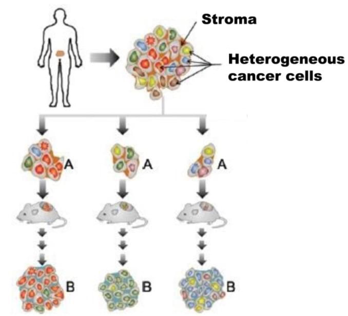
Generation of PDX-derived cells

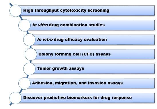
Applications

PDX-derived cell lines from Creative Bioarray are all early passage (<10) and maintain essential histopathological features and genetic profiles of the original patient tumors including biochemical signaling, genomic mutational status, and response to tumor cell autonomously targeted therapeutics. Each primary cell isolate consists of mouse stromal-cell-depleted primary cancer cells from a genetically defined PDX model, providing an excellent system to link in vitro to in vivo in the field of Oncology research.
Quotation and ordering
If you have any special needs in establishment or application of PDX-derived cell lines, please contact us for this special service. Let us know what you need and we will accommodate you. We look forward to working with you in the future.
References
- Lorin D,; Jennifer T,; et al. Primary esophageal and gastro-esophageal junction cancer xenograft models: clinicopathological features and engraftment. Laboratory Investigation. 2013, 93: 397–407.
- Natalie C,; Duncan I. J,; et al. Predictive in vivo animal models and translation to clinical trials. Drug Discovery Today. 2012, 17:253-260.
- Samuel AW,; et al. Patient-derived xenografts, the cancer stem cell paradigm, and cancer pathobiology in the 21st century. Laboratory Investigation. 2013, 93:970–982.
Explore Other Options
For research use only. Not for any other purpose.

