3D Angiogenesis Assay
- Service Details
- Features
- FAQ
- Explore Other Options
Angiogenesis is an incredibly intricate step in the development of a blood vessel, tightly controlled in space and time by pro- and anti-angiogenic molecules. This is important in both health and disease. Specifically, solid tumour growth and dissemination depends on angiogenesis, while other pathologies, such as atherosclerosis, are also directly dependent on angiogenesis. As a result, a variety of models have been developed to study the potential mechanisms of angiogenesis and their clinical applications. For in vitro studies, 3D modeling systems have irreplaceable advantages.
Why 3D angiogenesis?
- Angiogenesis in the tumor microenvironment is a multidimensional process, with complex interactions between tumor cells, stromal cells and capillaries in the microenvironment.
- In contrast to conventional 2D structures, 3D culture plates can mimic the structural and functional hallmarks of angiogenesis: endothelial cell migration, proliferation and lumen growth, branching and anastomosis.
- These in vitro 3D culture systems are useful both for the discovery of new drugs and the explanation of cancer drug resistance, and are suitable for high throughput analysis.
Creative Bioarray's angiogenesis assay utilizes a cutting-edge 3D model culture system that mimics in vivo angiogenesis and allows large-scale testing of test compounds for their pro- and anti-angiogenic activity on 96-well plates.
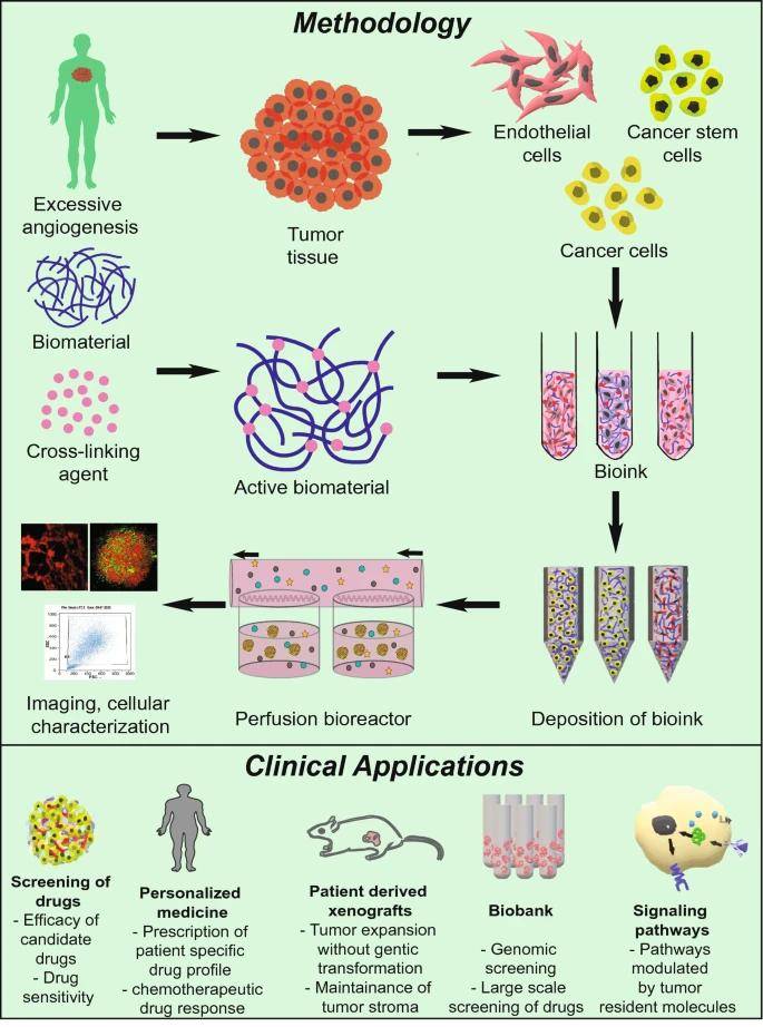 Fig. 1. Work flow to formulate 3D tumor vascular model systems and their clinical applications (Bhat S M., Badiger V A., et al., 2021).
Service Details
- 3D Medium Selection: We provide many types of 3D medium, not limited to the most well used Matrigel, Collagen I and Fibrin Gel, etc., for different research needs.
- Endothelial cell culture: We provide human umbilical vein endothelial cells (HUVEC) and other types of endothelial cells.
- Angiogenesis Stimulation: Add different concentrations of growth factors and cytokines as well as test compounds to stimulate or inhibit angiogenesis according to customers' needs.
- Assay Types:
Real-Time Tube Formation Assay: This assay monitors in real-time how endothelial cells are organized into capillaries.
Sprouting Assay: Used to measure endothelial sprouting from multicellular spheroids or matrix-encrusted chunks. - Data Collection and Analysis: Real-time monitoring of angiogenesis and comprehensive kinetic analysis using high-resolution microscopic imaging and multi-image readouts.
Service Workflow
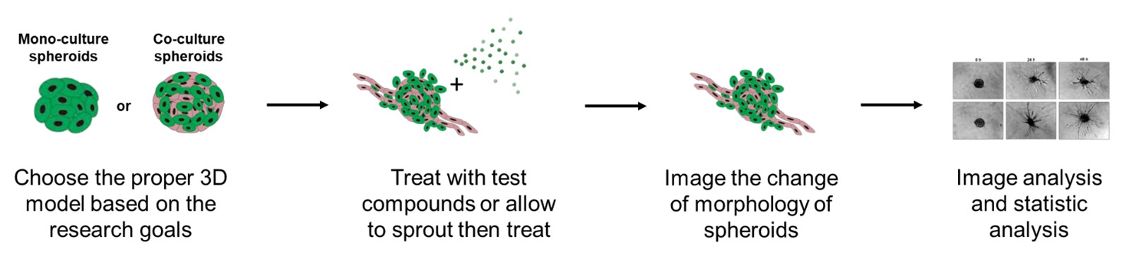 Fig. 2. 3D Angiogenesis Assay Workflow.
Advantage
- Select or customize the 3D culture system based on your needs and establish culture conditions that are close to the in vivo environment.
- Use multi-image readouts of the angiogenesis process to collect data including tube formation, branching patterns, and lumen development for comprehensive kinetic analysis.
- Test your compounds by choosing cells from our comprehensive human and animal cell bank or sending your own cells.
- Use models that capture the interaction between supporting cells and tumors cells.
- Multiple cell types can be considered and introduced into the 3D systems based on the bio-relevance of each tumor models.
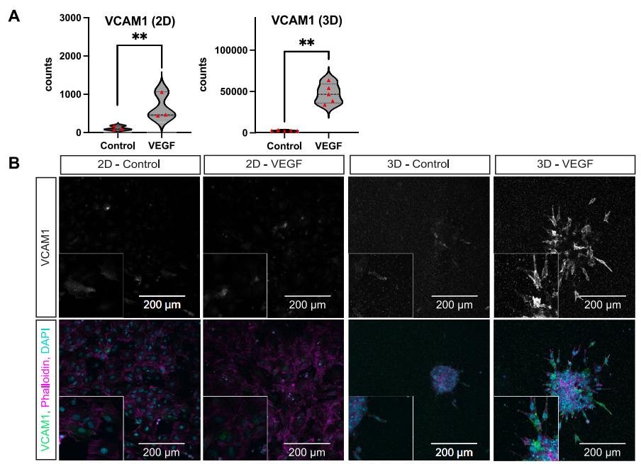 Fig. 3. Selected angiogenesis-associated genes are more prominently expressed and regulated in 3D settings on transcriptomic and protein level. (Rapp, J., Ness, J., et al., 2024)
FAQ
Q1: What is the difference between a 3D model and a 2D model?
The 3D angiogenesis assay allows you to more accurately model the 3D anatomy and cell microenvironment of tumor tissues, as well as the cell-cell interactions and the development of vascular networks, whereas 2D models are limited to a flat surface and are inadequate for studying more biological activity.
Q2: Which cells can be used in the assay?
Endothelial cells are our primary focus for angiogenesis, but we can scale the service to accommodate cell types you require for your specific research.
Q3: Does the 3D culture substrate used in the experiment comply with biosafety standards?
The 3D culture substrates we use comply with international biosafety standards to ensure the safety and reliability of the experiments.
Q4: Do you provide statistical analysis service for experimental data?
Yes, we provide professional statistical analysis services to help customers extract meaningful conclusions from experimental data.
Quotation and Ordering
If you have other questions, please contact us and we will provide you with detailed explanations. Our customer service representatives are available 24hr a day! Thanks for choosing Creative Bioarray services!
References
- Bhat SM, Badiger VA, et al. 3D tumor angiogenesis models: recent advances and challenges. J Cancer Res Clin Oncol. 2021. 147(12):3477-3494.
- Rapp, J., Ness, J., et al. 2D and 3D in vitro angiogenesis assays highlight different aspects of angiogenesis. Biochim Biophys Acta Mol Basis Dis. 2024 Mar;1870(3):167028.
Explore Other Options
