- You are here: Home
- Applications
- 3D Spheroid & Organoid Culture
- 3D Spheroid Platform for Drug Development
Applications
-
Cell Services
- Cell Line Authentication
- Cell Surface Marker Validation Service
-
Cell Line Testing and Assays
- Toxicology Assay
- Drug-Resistant Cell Models
- Cell Viability Assays
- Cell Proliferation Assays
- Cell Migration Assays
- Soft Agar Colony Formation Assay Service
- SRB Assay
- Cell Apoptosis Assays
- Cell Cycle Assays
- Cell Angiogenesis Assays
- DNA/RNA Extraction
- Custom Cell & Tissue Lysate Service
- Cellular Phosphorylation Assays
- Stability Testing
- Sterility Testing
- Endotoxin Detection and Removal
- Phagocytosis Assays
- Cell-Based Screening and Profiling Services
- 3D-Based Services
- Custom Cell Services
- Cell-based LNP Evaluation
-
Stem Cell Research
- iPSC Generation
- iPSC Characterization
-
iPSC Differentiation
- Neural Stem Cells Differentiation Service from iPSC
- Astrocyte Differentiation Service from iPSC
- Retinal Pigment Epithelium (RPE) Differentiation Service from iPSC
- Cardiomyocyte Differentiation Service from iPSC
- T Cell, NK Cell Differentiation Service from iPSC
- Hepatocyte Differentiation Service from iPSC
- Beta Cell Differentiation Service from iPSC
- Brain Organoid Differentiation Service from iPSC
- Cardiac Organoid Differentiation Service from iPSC
- Kidney Organoid Differentiation Service from iPSC
- GABAnergic Neuron Differentiation Service from iPSC
- Undifferentiated iPSC Detection
- iPSC Gene Editing
- iPSC Expanding Service
- MSC Services
- Stem Cell Assay Development and Screening
- Cell Immortalization
-
ISH/FISH Services
- In Situ Hybridization (ISH) & RNAscope Service
- Fluorescent In Situ Hybridization
- FISH Probe Design, Synthesis and Testing Service
-
FISH Applications
- Multicolor FISH (M-FISH) Analysis
- Chromosome Analysis of ES and iPS Cells
- RNA FISH in Plant Service
- Mouse Model and PDX Analysis (FISH)
- Cell Transplantation Analysis (FISH)
- In Situ Detection of CAR-T Cells & Oncolytic Viruses
- CAR-T/CAR-NK Target Assessment Service (ISH)
- ImmunoFISH Analysis (FISH+IHC)
- Splice Variant Analysis (FISH)
- Telomere Length Analysis (Q-FISH)
- Telomere Length Analysis (qPCR assay)
- FISH Analysis of Microorganisms
- Neoplasms FISH Analysis
- CARD-FISH for Environmental Microorganisms (FISH)
- FISH Quality Control Services
- QuantiGene Plex Assay
- Circulating Tumor Cell (CTC) FISH
- mtRNA Analysis (FISH)
- In Situ Detection of Chemokines/Cytokines
- In Situ Detection of Virus
- Transgene Mapping (FISH)
- Transgene Mapping (Locus Amplification & Sequencing)
- Stable Cell Line Genetic Stability Testing
- Genetic Stability Testing (Locus Amplification & Sequencing + ddPCR)
- Clonality Analysis Service (FISH)
- Karyotyping (G-banded) Service
- Animal Chromosome Analysis (G-banded) Service
- AAV Biodistribution Analysis (RNA ISH)
- Molecular Karyotyping (aCGH)
- Droplet Digital PCR (ddPCR) Service
- Digital ISH Image Quantification and Statistical Analysis
- SCE (Sister Chromatid Exchange) Analysis
- Biosample Services
- Histology Services
- Exosome Research Services
- In Vitro DMPK Services
-
In Vivo DMPK Services
- Pharmacokinetic and Toxicokinetic
- PK/PD Biomarker Analysis
- Bioavailability and Bioequivalence
- Bioanalytical Package
- Metabolite Profiling and Identification
- In Vivo Toxicity Study
- Mass Balance, Excretion and Expired Air Collection
- Administration Routes and Biofluid Sampling
- Quantitative Tissue Distribution
- Target Tissue Exposure
- In Vivo Blood-Brain-Barrier Assay
- Drug Toxicity Services
3D Spheroid Platform for Drug Development
3D spheroids are formed by culturing cells in a three-dimensional environment, enabling them to interact and form complex structures like those found in vivo. In 3D multicellular spheroids, metabolic gradients can form, resulting in a diverse mix of cells that interact exceptionally well with each other and the extracellular matrix. Compared to traditional 2D cell culture, 3D spheroids provide a more realistic representation of the body's environment and effectively mimic various tissue types in diseased conditions, enhancing clarity and fluidity.
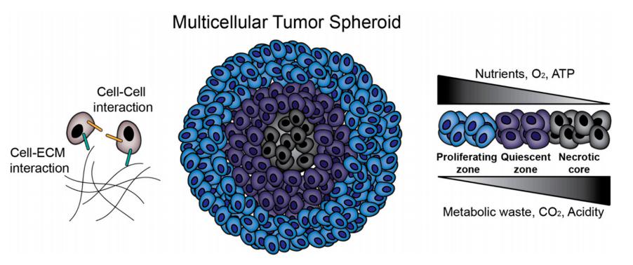
Figure 1. Multicellular tumor spheroids (MCTS) biology.[1]
3D spheroids have emerged as a powerful tool in drug development, offering significant advantages over traditional 2D cell culture systems. These spherical aggregates of cells better mimic the in vivo tumor microenvironment, providing insights into cellular behavior, drug responses, and the overall efficacy and safety of therapeutic compounds.
Features
- Structural Relevance
3D spheroids maintain a more physiologically relevant architecture compared to 2D cultures. They can better replicate cell-cell and cell-matrix interactions, which are crucial for cellular functions.
- Microenvironment Simulation
The internal gradients of nutrients, oxygen, and pH in 3D spheroids mimic those found in solid tumors, making them particularly useful for studying tumor biology and drug response.
- Cell-typical Behavior
Cells in 3D cultures often exhibit differentiated phenotypes and behaviors, such as altered gene expression and increased resistance to drugs, closely resembling the in vivo situation.
3D Spheroid Models Applications
- Cancer Research
3D spheroids are used to model tumor growth and metastasis. They provide a more accurate representation of tumor microenvironments compared to traditional 2D cultures, allowing researchers to study drug responses, cellular behavior, and treatment efficacy.
- Drug Development and Screening
Spheroids provide a more predictive platform for testing the efficacy and toxicity of new pharmaceuticals. They mimic the in vivo environment better than 2D cultures, helping to identify potential drug candidates early in the development process.
- Toxicology Testing
Spheroids can be used to evaluate the toxicological effects of chemicals and drugs, providing insights into safety and efficacy before clinical trials.
- Migration and Invasion Assays
Cancer cells in 3D spheroids display behaviors more similar to those observed in tumors, including heterogeneity and phenotypic variations, which are crucial for studying the biology of cancer cell migration and invasion.
Creative Bioarray provides advanced 3D spheroid models designed for researchers aiming to explore cell behavior in a more realistic environment. Our 3D Spheroid for Drug Development enables in-depth investigation of cell-cell interactions, drug responses, and cell migration, offering enhanced physiological relevance for your studies.

Reference
- Kim, Sungjin et al. "Uniform sized cancer spheroids production using hydrogel-based droplet microfluidics: a review." Biomedical microdevices vol. 26,2 26. 29 May. 2024, doi:10.1007/s10544-024-00712-3
Explore Other Options
For research use only. Not for any other purpose.