- You are here: Home
- Services
- Cell Services
- 3D-Based Services
- 3D Invasion Assay
Services
-
Cell Services
- Cell Line Authentication
- Cell Surface Marker Validation Service
-
Cell Line Testing and Assays
- Toxicology Assay
- Drug-Resistant Cell Models
- Cell Viability Assays
- Cell Proliferation Assays
- Cell Migration Assays
- Soft Agar Colony Formation Assay Service
- SRB Assay
- Cell Apoptosis Assays
- Cell Cycle Assays
- Cell Angiogenesis Assays
- DNA/RNA Extraction
- Custom Cell & Tissue Lysate Service
- Cellular Phosphorylation Assays
- Stability Testing
- Sterility Testing
- Endotoxin Detection and Removal
- Phagocytosis Assays
- Cell-Based Screening and Profiling Services
- 3D-Based Services
- Custom Cell Services
- Cell-based LNP Evaluation
-
Stem Cell Research
- iPSC Generation
- iPSC Characterization
-
iPSC Differentiation
- Neural Stem Cells Differentiation Service from iPSC
- Astrocyte Differentiation Service from iPSC
- Retinal Pigment Epithelium (RPE) Differentiation Service from iPSC
- Cardiomyocyte Differentiation Service from iPSC
- T Cell, NK Cell Differentiation Service from iPSC
- Hepatocyte Differentiation Service from iPSC
- Beta Cell Differentiation Service from iPSC
- Brain Organoid Differentiation Service from iPSC
- Cardiac Organoid Differentiation Service from iPSC
- Kidney Organoid Differentiation Service from iPSC
- GABAnergic Neuron Differentiation Service from iPSC
- Undifferentiated iPSC Detection
- iPSC Gene Editing
- iPSC Expanding Service
- MSC Services
- Stem Cell Assay Development and Screening
- Cell Immortalization
-
ISH/FISH Services
- In Situ Hybridization (ISH) & RNAscope Service
- Fluorescent In Situ Hybridization
- FISH Probe Design, Synthesis and Testing Service
-
FISH Applications
- Multicolor FISH (M-FISH) Analysis
- Chromosome Analysis of ES and iPS Cells
- RNA FISH in Plant Service
- Mouse Model and PDX Analysis (FISH)
- Cell Transplantation Analysis (FISH)
- In Situ Detection of CAR-T Cells & Oncolytic Viruses
- CAR-T/CAR-NK Target Assessment Service (ISH)
- ImmunoFISH Analysis (FISH+IHC)
- Splice Variant Analysis (FISH)
- Telomere Length Analysis (Q-FISH)
- Telomere Length Analysis (qPCR assay)
- FISH Analysis of Microorganisms
- Neoplasms FISH Analysis
- CARD-FISH for Environmental Microorganisms (FISH)
- FISH Quality Control Services
- QuantiGene Plex Assay
- Circulating Tumor Cell (CTC) FISH
- mtRNA Analysis (FISH)
- In Situ Detection of Chemokines/Cytokines
- In Situ Detection of Virus
- Transgene Mapping (FISH)
- Transgene Mapping (Locus Amplification & Sequencing)
- Stable Cell Line Genetic Stability Testing
- Genetic Stability Testing (Locus Amplification & Sequencing + ddPCR)
- Clonality Analysis Service (FISH)
- Karyotyping (G-banded) Service
- Animal Chromosome Analysis (G-banded) Service
- I-FISH Service
- AAV Biodistribution Analysis (RNA ISH)
- Molecular Karyotyping (aCGH)
- Droplet Digital PCR (ddPCR) Service
- Digital ISH Image Quantification and Statistical Analysis
- SCE (Sister Chromatid Exchange) Analysis
- Biosample Services
- Histology Services
- Exosome Research Services
- In Vitro DMPK Services
-
In Vivo DMPK Services
- Pharmacokinetic and Toxicokinetic
- PK/PD Biomarker Analysis
- Bioavailability and Bioequivalence
- Bioanalytical Package
- Metabolite Profiling and Identification
- In Vivo Toxicity Study
- Mass Balance, Excretion and Expired Air Collection
- Administration Routes and Biofluid Sampling
- Quantitative Tissue Distribution
- Target Tissue Exposure
- In Vivo Blood-Brain-Barrier Assay
- Drug Toxicity Services
3D Invasion Assay

Invasion and metastasis are responsible for the high morbidity and mortality in cancer patients. Invasion into the surrounding normal tissues is generally considered to be a key hallmark of malignant tumors. More efforts for anticancer drug development now move towards therapies that targeted on the inhibition of this key "hallmark" of cancer. The 3D tumor spheroid invasion assay allows cells to invade out of the tumor mass into the extracellular matrix-like environment. The true 3D manner takes into account important aspects of the pathophysiology of a tumor mass, in which the tumor spheroids may experience hypoxia and nutrient deprivation which may cause the change in gene expression and promote migration and invasion. Creative Bioarray believes that the rapid, automatable 3D invasion system that enables highly reproducible experimental conditions and high throughput analysis will give our customers the best results to identify novel therapeutic agents.
Creative Bioarray 3D invasion assay service advantages:
- Test your compounds by choosing cells from our comprehensive human and animal cell bank or sending your own cells.
- Set the suitable surrounding matrix according to different biological relevance of the tumor models.
- High-throughput, quantitative, and real time detection.
- Rapid turnaround time
Principle
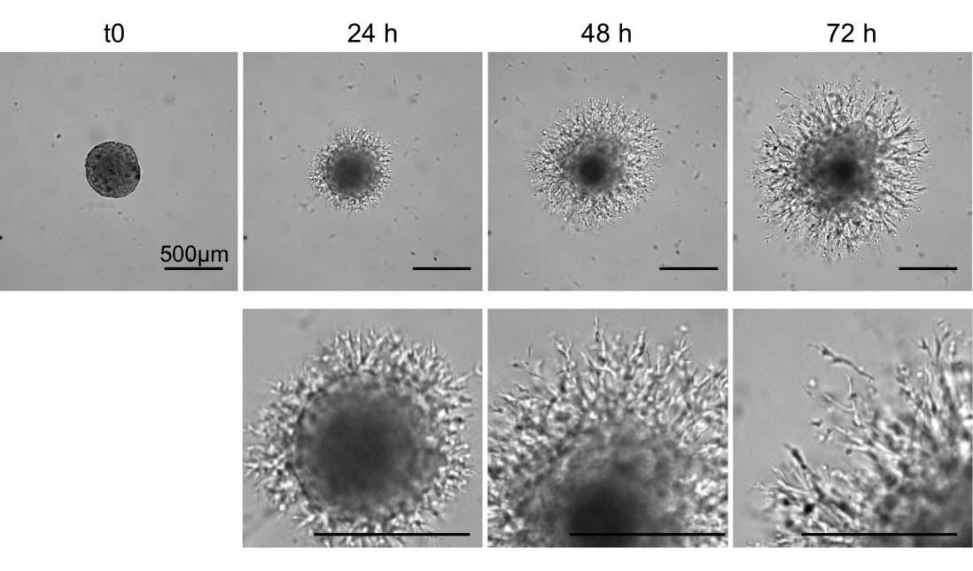
3D tumor spheroids invasion assays: Multicellular spheroids are embedded into 3D ECM. It is expected to see that non-invasive cancer cells stay as compact spheroids with a distinct border to the surrounding ECM and do not show any obvious signs of invasion. However, invasive cells or endothelial cells start to invade into the surrounding matrix and display outgrowth from the spheroids.
Workflow

Applications
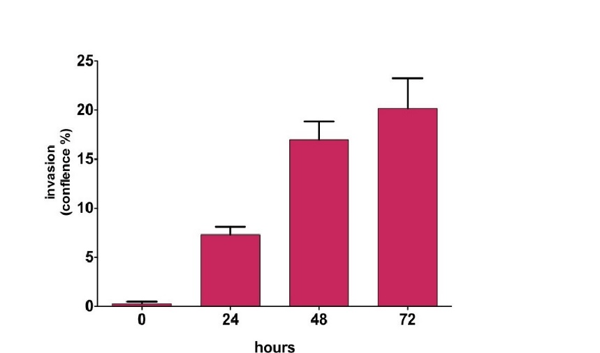
- The live images can be quantified for the invasive area over time. The invadopodia extending into the ECM will be recorded by the measurement of different parameters (area, diameter, perimeter etc.
- Different substrates for 3D microenvironment are available in Creative Bioarray depending on the research questions.
- The interactions between cancer spheroids and other cell types (i.e., stromal cells) can be captured be these assays. Dispersed stromal cells in ECM in addition to cancer spheroids may activate the invasive properties and other molecular functions.
Study examples
 Figure 1 The representative images for 3D invasion assay for glioblastoma spheroids
 Figure 2 The representative image analysis of 3D invasion assay for glioblastoma spheroids
Quotations and ordering
Our customer service representatives are available 24hr a day!
We also provide 2D cell migration and invasion service for our customers.
References
- Vinci, M., et al. Advances in establishment and analysis of three-dimensional tumor spheroid-based functional assays for target validation and drug evaluation. BMC biology. 2012, 10.1: 29.
- Kramer, N., et al. In vitro cell migration and invasion assays. Mutation Research/Reviews in Mutation Research. 2013, 752.1: 10-24.
- Vinci, M M., et al. Three-dimensional (3D) tumor spheroid invasion assay. JoVE (Journal of Visualized Experiments). 2015, 99: e52686-e52686.
Explore Other Options
For research use only. Not for any other purpose.

