Prostate Tumor Cells
- Background
- Applications
- Scientific Data
Prostate tumor cells usually arise from the prostatic epithelium, specifically from the cells lining the prostate ducts. These cells can undergo malignant transformation due to a variety of factors, including genetic mutations, hormonal influences, and environmental exposures. Some prostate cancers remain localized to the gland and may not cause significant harm, while others can invade nearby tissues and spread to other parts of the body, such as the bones and lymph nodes, a process known as metastasis.
Characteristics of Prostate Tumor Cells
- Cellular architecture and morphology. Prostate tumor cells exhibit distinct morphological features when compared to normal prostate cells. These tumor cells often display irregular shapes, increased nuclear-to-cytoplasmic ratios, and heterogeneous chromatin patterns. Such features are commonly assessed in histopathological examinations, leading to classification according to the Gleason grading system, which aids in predicting clinical outcomes.
- Genetic alterations. Prostate tumor cells frequently harbor key genetic mutations that contribute to tumorigenesis. Notable among these are alterations in the TP53 and PTEN tumor suppressor genes, which disrupt critical cellular regulatory pathways. Additionally, mutations in the AR gene, responsible for androgen receptor function, are linked to advanced disease and treatment resistance.
Research and Experimental Models
Prostate tumor cells are instrumental in preclinical research, providing insights into tumor biology and treatment efficacy. Utilizing cell lines and patient-derived xenografts allows researchers to study the cellular response to various therapeutics in a controlled environment. Such models are essential for expanding the understanding of the tumor microenvironment and the cellular interactions that drive prostate cancer progression.
Therapeutic Targeting
Prostate tumor cells provide a valuable model for exploring targeted therapeutic approaches. Recently, the development of next-generation inhibitors, such as PARP inhibitors for BRCA-mutated tumors, has gained traction. These therapeutics aim to exploit specific vulnerabilities of prostate tumor cells, thereby improving patient outcomes. Furthermore, immunotherapeutic strategies, including prostate-specific antigen (PSA) vaccines, aim to harness the patient's immune system to target and eliminate tumor cells effectively.
rADAM9D Inhibits the Invasion and Migration of DU145 Prostate Tumor Cells
The ADAM (A Disintegrin And Metalloproteinase) family of proteins comprises a group of multifunctional proteins that play important roles in many biological processes, such as cell fusion, cell adhesion, proteolysis, and in some diseases as well, including cancer. rADAM9D was tested for its ability to inhibit DU145 cell invasion and migration. rADAM9D, at concentrations ranging from 100 to 1000 nM, was able to significantly inhibit DU145 cell invasion (Fig. 1). In a wound healing migration assay, rADAM9D significantly inhibited DU145 cell migration at 100, 500, 1000, and 2000nM (Fig. 2A and B). The effects were more striking after 24 hours of wound repopulation.
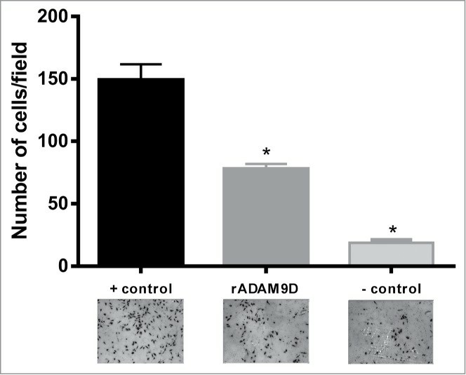 Fig.
1 rADAM9D inhibits the invasion of DU145 cells through matrigel. (Martin AC, et al, 2015)
Fig.
1 rADAM9D inhibits the invasion of DU145 cells through matrigel. (Martin AC, et al, 2015)
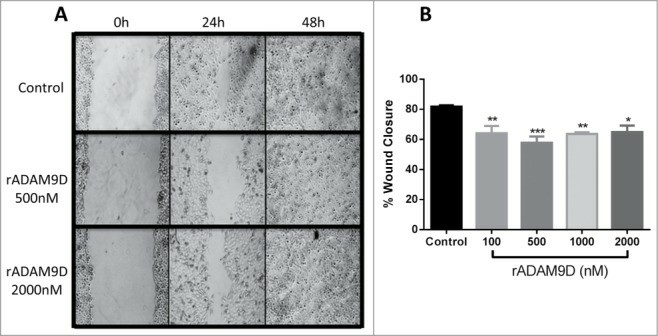 Fig. 2 rADAM9D inhibits the
migration of DU145 cells in a wound healing assay. (A) Cells were incubated with ADAM9D (100, 500, 1000, and
2000 nM) for 24 h and 48 h. Only pictures from rADAMD9 500 and 2000 nM are represented. (B) The closure area of
migrating cells was measured using ImageJ software, and it was calculated the percentage of wound closure,
comparing time zero and 24 hours. Results are expressed as percent of wound closure relative to control
(untreated) cells. (Martin AC, et al, 2015)
Fig. 2 rADAM9D inhibits the
migration of DU145 cells in a wound healing assay. (A) Cells were incubated with ADAM9D (100, 500, 1000, and
2000 nM) for 24 h and 48 h. Only pictures from rADAMD9 500 and 2000 nM are represented. (B) The closure area of
migrating cells was measured using ImageJ software, and it was calculated the percentage of wound closure,
comparing time zero and 24 hours. Results are expressed as percent of wound closure relative to control
(untreated) cells. (Martin AC, et al, 2015)
Ad Efficiently Transduce Human and Mouse Prostate Cancer Cells in Vitro
Prostate diseases are common in males worldwide with high morbidity. Gene therapy is an attractive therapeutic strategy for prostate diseases, however, it is currently underdeveloped. As well known, adenovirus (Ad) is the most widely used gene therapy vector. To evaluate the transduction efficiency of Ad in prostate cancer cells in vitro, PC3 and TRAMP-C2 cells, a human and mouse prostate cancer cell line, respectively, were infected with indicated MOI of EGFP-expressing Ad. One day post-infection, the transduction efficiency of Ad was quantitatively profiled by measuring the overall EGFP fluorescence intensity (Fig. 3A, B) and the percentage of EGFP-positive cells (Fig. 3C), respectively. The data showed that Ad transduced TRAMP-C2 cells consistently better than PC3 cells with MOI ranging from 1 to 105 (Fig. 3B, C). Taken together, Ad could efficiently transduce human and mouse prostate cancer cells in vitro in a dose-dependent manner.
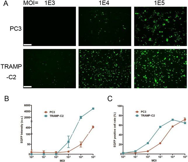 Fig. 3 (A) Representative
microscopic images showing EGFP fluorescence signal of PC3 and TRAMP-C2 cells infected with indicated MOI of
EGFP-expressing Ad vectors. (B) Quantification of EGFP intensity presented in arbitrary unit (a.u.). (C)
Percentage of EGFP-positive cells analyzed by flow cytometry. (Ai J, et al., 2017)
Fig. 3 (A) Representative
microscopic images showing EGFP fluorescence signal of PC3 and TRAMP-C2 cells infected with indicated MOI of
EGFP-expressing Ad vectors. (B) Quantification of EGFP intensity presented in arbitrary unit (a.u.). (C)
Percentage of EGFP-positive cells analyzed by flow cytometry. (Ai J, et al., 2017)
Filters Clear all filters
Species
- Cat (1)
- Human (992)
- Mouse (5)
- Rat (1)
Source
- Abdomen Metastasis (2)
- Adrenal Gland (7)
- Adrenal Gland Metastasis (2)
- Ascites (23)
- Ascites Metastasis (32)
- Bile Duct (3)
- Bladder (12)
- Blood (120)
- Bone (21)
- Bone Marrow (43)
- Bone Marrow Metastasis (18)
- Bone Metastasis (6)
- Brain (31)
- Brain Metastasis (6)
- Breast (8)
- Bronchus (1)
- Cecum (3)
- Cerebrospinal Fluid (1)
- Cerebrospinal Fluid Metastasis (1)
- Cervix (32)
- Colon (84)
- Cornea (3)
- Cutaneous Metastasis (1)
- Dermis (1)
- Duodenum (1)
- Endometrium (17)
- Esophagus (44)
- Eye (12)
- Eye Socket (5)
- Fetus (1)
- Foreskin (4)
- Gallbladder (1)
- Gingiva (2)
- Globe (2)
- Groin (1)
- Hypodermis Metastasis (5)
- Intestine (85)
- kidney (1)
- Kidney (9)
- Liver (13)
- Liver Metastasis (17)
- Lung (42)
- Lung Metastasis (8)
- Lymph Node (5)
- Lymph Node Metastasis (58)
- Muscle (4)
- Muscle Metastasis (2)
- Nose (2)
- Omentum Metastasis (2)
- Oral Cavity (10)
- Ovary (13)
- Ovary Metastasis (2)
- Pancreas (10)
- Pelvic Wall Metastasis (1)
- Pelvis (1)
- Perianal Space Metastasis (1)
- Pericardial Effusion (1)
- Pericardial Effusion Metastasis (1)
- Perineus (1)
- Peripheral Blood (119)
- Peritoneal Effusion (2)
- Peritoneum (1)
- Peritoneum Metastasis (1)
- Pharynx (3)
- Pleural Effusion (54)
- Pleural Effusion Metastasis (44)
- Prostate (4)
- Rectum (13)
- Renal Pelvis (1)
- Retroperitoneal Space (2)
- Salivary Gland (2)
- Skeletal Muscle (1)
- Skin (22)
- Skin Metastasis (3)
- Small Intestine (1)
- Small Intestine Metastasis (1)
- Soft Tissue Metastasis (1)
- Stomach (4)
- Testis (9)
- Thoracic Cavity Metastasis (6)
- Thyroid Gland (15)
- Thyroid Gland Metastasis (1)
- Tongue (5)
- Umbilical Cord (1)
- Umbilical Cord Blood (1)
- Urachus (1)
- Ureter (1)
- Uterus (53)
- Uvea (2)
- Vagina (2)
- Vulva (1)
Disease
- Acute Biphenotypic Leukemia (1)
- Acute Erythroid Leukemia (4)
- Acute Megakaryoblastic Leukemia (4)
- Acute Monocytic Leukemia (9)
- Acute Myeloid Leukemia (25)
- Acute Promyelocytic Leukemia (2)
- Adrenal Gland Neuroblastoma (11)
- Adult B Acute Lymphoblastic leukemia (1)
- Adult B Acute Lymphoblastic Leukemia (6)
- Adult T Acute Lymphoblastic Leukemia (6)
- Adult T Lymphoblastic Lymphoma (2)
- Adult T-Cell Leukemia/Lymphoma (1)
- Alveolar Rhabdomyosarcoma (4)
- Alveolar Ridge Squamous Cell Carcinoma (1)
- Amelanotic Melanoma (3)
- Ampulla of Vater Adenocarcinoma (1)
- Ampulla of Vater Adenosquamous Carcinoma (3)
- Anaplastic Astrocytoma (3)
- Anaplastic Large Cell Lymphoma (7)
- Askin Tumor (1)
- Astrocytoma (5)
- B Acute Lymphoblastic Leukemia (2)
- B-Cell Non-Hodgkin Lymphoma (5)
- Bare Lymphocyte Syndrome Type 2 (1)
- Barrett Adenocarcinoma (2)
- Benign Prostatic Hyperplasia (1)
- Bladder Carcinoma (12)
- Bladder Squamous Cell Carcinoma (1)
- Breast Adenocarcinoma (1)
- Breast Carcinoma (9)
- Breast Ductal Carcinoma (2)
- Burkitt Lymphoma (17)
- Canavan Disease (1)
- Cecum Adenocarcinoma (3)
- Central Nervous System Lymphoma (2)
- Cervical Adenocarcinoma (2)
- Cervical Adenosquamous Carcinoma (2)
- Cervical Small Cell Carcinoma (1)
- Cervical Squamous Cell Carcinoma (2)
- Childhood B Acute Lymphoblastic Leukemia (13)
- Childhood T Acute Lymphoblastic Leukemia (16)
- Childhood T Lymphoblastic Lymphoma (1)
- Cholangiocarcinoma (2)
- Chronic Eosinophilic Leukemia (1)
- Chronic Lymphocytic Leukemia (2)
- Chronic Myeloid Leukemia (23)
- Clear Cell Renal Cell Carcinoma (2)
- Colon Adenocarcinoma (55)
- Colon Carcinoma (34)
- Colorectal Adenocarcinoma (1)
- Colorectal Carcinoma (1)
- Congenital Pure Red Cell Aplasia (1)
- Cutaneous Melanoma (10)
- Dedifferentiated Chondrosarcoma (1)
- Desmoplastic Melanoma (1)
- Diffuse Large B-Cell Lymphoma (28)
- Down Syndrome (2)
- EBV-Related Burkitt Lymphoma (12)
- Embryonal Carcinoma (3)
- Embryonal Rhabdomyosarcoma (3)
- Endometrial Adenocarcinoma (13)
- Endometrial Adenosquamous Carcinoma (2)
- Endometrial Carcinoma (2)
- Endometrioid Stromal Sarcoma (1)
- Epithelioid Hemangioendothelioma (1)
- Epithelioid Sarcoma (3)
- Esophageal Adenocarcinoma (6)
- Esophageal Squamous Cell Carcinoma (41)
- Essential Thrombocythemia (1)
- Ewing Sarcoma (2)
- Extraskeletal Myxoid Chondrosarcoma (1)
- Fanconi Anemia (1)
- Fibrosarcoma (1)
- Follicular Lymphoma (2)
- Gallbladder Carcinoma (2)
- Gallbladder Undifferentiated Carcinoma (2)
- Gastric Adenocarcinoma (6)
- Gastric Adenosquamous Carcinoma (1)
- Gastric Carcinoma (5)
- Gastric Choriocarcinoma (1)
- Gastric Fundus Carcinoma (1)
- Gastric Signet Ring Cell Adenocarcinoma (1)
- Gastric Small Cell Carcinoma (2)
- Gastric Tubular Adenocarcinoma (5)
- Gastroesophageal Junction Adenocarcinoma (1)
- Gestational Choriocarcinoma (1)
- Gingival Squamous Cell Carcinoma (2)
- Glioblastoma (18)
- Gliosarcoma (1)
- Hairy Cell Leukemia (1)
- Hepatoblastoma (2)
- Hepatocellular Carcinoma (6)
- Hepatosplenic T-Cell Lymphoma (2)
- Hereditary Thyroid Gland Medullary Carcinoma (1)
- High Grade B-Cell Lymphoma (1)
- High Grade Ovarian Serous Adenocarcinoma (8)
- Hodgkin Lymphoma (9)
- Hypopharyngeal Squamous Cell Carcinoma (2)
- Infectious Mononucleosis (1)
- Intrahepatic Cholangiocarcinoma (6)
- Invasive Breast Carcinoma of No Special Type (12)
- Kidney Neoplasm (1)
- Kidney Rhabdoid Tumor (1)
- Krukenberg Tumor (1)
- Liposarcoma (1)
- Lung Adenocarcinoma (17)
- Lung Giant Cell Carcinoma (8)
- Lung Large Cell Carcinoma (9)
- Lung Mucoepidermoid Carcinoma (1)
- Lung Non-Small Cell Carcinoma (2)
- Lung Small Cell Carcinoma (25)
- Lung Squamous Cell Carcinoma (9)
- Lymphoblastic Lymphoma (1)
- Malignant Peripheral Nerve Sheath Tumor (1)
- Mantle Cell Lymphoma (5)
- Mature Gastric Teratoma (1)
- Maxillary Sinus Squamous Cell Carcinoma (1)
- Medulloblastoma (3)
- Melanoma (24)
- Meningioma (2)
- Minimally Invasive Lung Adenocarcinoma (1)
- Monophasic Synovial Sarcoma (1)
- Mouse Intestinal Tract Neuroendocrine Adenoma (1)
- Mouse Mammary Gland Malignant Neoplasm (2)
- Mouse Plasmacytoma (1)
- Mycosis Fungoides (1)
- Myelodysplastic Syndrome (1)
- Myxofibrosarcoma (1)
- Natural Killer Cell Lymphoblastic Leukemia/Lymphoma (2)
- Neuroblastoma (26)
- Oral Cavity Squamous Cell Carcinoma (15)
- Osteoid Osteoma (1)
- Osteosarcoma (15)
- Ovarian Carcinoma (1)
- Ovarian Clear Cell Adenocarcinoma (1)
- Ovarian Endometrioid Adenocarcinoma (4)
- Ovarian Granulosa Cell Tumor (1)
- Ovarian Mucinous Adenocarcinoma (2)
- Ovarian Serous Adenocarcinoma (2)
- Ovarian Serous Cystadenocarcinoma (2)
- Ovarian Small Cell Carcinoma (1)
- Pancreatic Adenocarcinoma (13)
- Pancreatic Carcinoma (5)
- Pancreatic Ductal Adenocarcinoma (12)
- Papillomavirus-Independent Cervical Squamous Cell Carcinoma (1)
- Papillomavirus-Related Cervical Adenocarcinoma (7)
- Papillomavirus-Related Cervical Squamous Cell Carcinoma (4)
- Papillomavirus-Related Endocervical Adenocarcinoma (16)
- Paroxysmal Nocturnal Hemoglobinuria (3)
- Pharyngeal Squamous Cell Carcinoma (1)
- Plasma Cell Myeloma (15)
- Pleural Epithelioid Mesothelioma (5)
- Pleural Sarcomatoid Mesothelioma (2)
- Poorly Differentiated Thyroid Gland Carcinoma (1)
- Primary Cutaneous T-Cell Non-Hodgkin Lymphoma (1)
- Primary Effusion Lymphoma (7)
- Primitive Neuroectodermal Tumor (1)
- Prostate carcinoma (1)
- Prostate Carcinoma (9)
- Rectal Adenocarcinoma (13)
- Rectosigmoid Adenocarcinoma (1)
- Recurrent Bladder Carcinoma (1)
- Renal Cell Carcinoma (7)
- Renal Pelvis Urothelial Carcinoma (1)
- Retinoblastoma (11)
- Sacral Chordoma (1)
- Sacrococcygeal Teratoma (1)
- Salivary Gland Squamous Cell Carcinoma (1)
- Sezary Syndrome (1)
- Shwachman-Diamond Syndrome (1)
- Skin Squamous Cell Carcinoma (2)
- Splenic Marginal Zone Lymphoma (1)
- Testicular Embryonal Carcinoma (8)
- Testicular Teratoma (2)
- Testicular Yolk Sac Tumor (1)
- Thyroid Gland Anaplastic Carcinoma (10)
- Thyroid Gland Follicular Carcinoma (4)
- Thyroid Gland Papillary Carcinoma (3)
- Thyroid Gland Sarcoma (1)
- Thyroid Gland Squamous Cell Carcinoma (2)
- Tongue Adenosquamous Carcinoma (1)
- Tongue Squamous Cell Carcinoma (6)
- Type I Endometrial Adenocarcinoma (1)
- Ureter Urothelial Carcinoma (1)
- Uterine Carcinosarcoma (2)
- Uterine Corpus Leiomyosarcoma (1)
- Uterine Corpus Sarcoma (2)
- Uveal Melanoma (2)
- Vaginal Melanoma (2)
- Vulvar Melanoma (1)
- Vulvar Squamous Cell Carcinoma (1)

Description: prostate epithelial cells from a 68-year-old man with benign prostate hyperplasia; cells were ...

Description: Cytogenetic analysis and DNA fingerprinting at the Creative Bioarray documented that this cell line ...

Description: Human cell line derived from malignant peripheral nerve sheath tumor.
Description: C4-2 is a cell line with epithelial-like morphology that was isolated from a human prostate cancer ...

Description: This cell line was established from prostate cancer tissue harvested from a metastatic lesion to a ...

Description: Derived from a human prostate carcinoma xenograft (CWR22R) that was serially propagated in nude ...
Description: 22Rv1 is a human prostate carcinoma epithelial cell line derived from a xenograft that was serially ...

Description: These cells are responsive to 5-alpha-dihydrotestosterone (growth modulation and acid phosphatase); ...