- You are here: Home
- Services
- Cell Services
- Cell Line Testing and Assays
- Sulforhodamine B (SRB) Assay
Services
-
Cell Services
- Cell Line Authentication
- Cell Surface Marker Validation Service
-
Cell Line Testing and Assays
- Toxicology Assay
- Drug-Resistant Cell Models
- Cell Viability Assays
- Cell Proliferation Assays
- Cell Migration Assays
- Soft Agar Colony Formation Assay Service
- SRB Assay
- Cell Apoptosis Assays
- Cell Cycle Assays
- Cell Angiogenesis Assays
- DNA/RNA Extraction
- Custom Cell & Tissue Lysate Service
- Cellular Phosphorylation Assays
- Stability Testing
- Sterility Testing
- Endotoxin Detection and Removal
- Phagocytosis Assays
- Cell-Based Screening and Profiling Services
- 3D-Based Services
- Custom Cell Services
- Cell-based LNP Evaluation
-
Stem Cell Research
- iPSC Generation
- iPSC Characterization
-
iPSC Differentiation
- Neural Stem Cells Differentiation Service from iPSC
- Astrocyte Differentiation Service from iPSC
- Retinal Pigment Epithelium (RPE) Differentiation Service from iPSC
- Cardiomyocyte Differentiation Service from iPSC
- T Cell, NK Cell Differentiation Service from iPSC
- Hepatocyte Differentiation Service from iPSC
- Beta Cell Differentiation Service from iPSC
- Brain Organoid Differentiation Service from iPSC
- Cardiac Organoid Differentiation Service from iPSC
- Kidney Organoid Differentiation Service from iPSC
- GABAnergic Neuron Differentiation Service from iPSC
- Undifferentiated iPSC Detection
- iPSC Gene Editing
- iPSC Expanding Service
- MSC Services
- Stem Cell Assay Development and Screening
- Cell Immortalization
-
ISH/FISH Services
- In Situ Hybridization (ISH) & RNAscope Service
- Fluorescent In Situ Hybridization
- FISH Probe Design, Synthesis and Testing Service
-
FISH Applications
- Multicolor FISH (M-FISH) Analysis
- Chromosome Analysis of ES and iPS Cells
- RNA FISH in Plant Service
- Mouse Model and PDX Analysis (FISH)
- Cell Transplantation Analysis (FISH)
- In Situ Detection of CAR-T Cells & Oncolytic Viruses
- CAR-T/CAR-NK Target Assessment Service (ISH)
- ImmunoFISH Analysis (FISH+IHC)
- Splice Variant Analysis (FISH)
- Telomere Length Analysis (Q-FISH)
- Telomere Length Analysis (qPCR assay)
- FISH Analysis of Microorganisms
- Neoplasms FISH Analysis
- CARD-FISH for Environmental Microorganisms (FISH)
- FISH Quality Control Services
- QuantiGene Plex Assay
- Circulating Tumor Cell (CTC) FISH
- mtRNA Analysis (FISH)
- In Situ Detection of Chemokines/Cytokines
- In Situ Detection of Virus
- Transgene Mapping (FISH)
- Transgene Mapping (Locus Amplification & Sequencing)
- Stable Cell Line Genetic Stability Testing
- Genetic Stability Testing (Locus Amplification & Sequencing + ddPCR)
- Clonality Analysis Service (FISH)
- Karyotyping (G-banded) Service
- Animal Chromosome Analysis (G-banded) Service
- I-FISH Service
- AAV Biodistribution Analysis (RNA ISH)
- Molecular Karyotyping (aCGH)
- Droplet Digital PCR (ddPCR) Service
- Digital ISH Image Quantification and Statistical Analysis
- SCE (Sister Chromatid Exchange) Analysis
- Biosample Services
- Histology Services
- Exosome Research Services
- In Vitro DMPK Services
-
In Vivo DMPK Services
- Pharmacokinetic and Toxicokinetic
- PK/PD Biomarker Analysis
- Bioavailability and Bioequivalence
- Bioanalytical Package
- Metabolite Profiling and Identification
- In Vivo Toxicity Study
- Mass Balance, Excretion and Expired Air Collection
- Administration Routes and Biofluid Sampling
- Quantitative Tissue Distribution
- Target Tissue Exposure
- In Vivo Blood-Brain-Barrier Assay
- Drug Toxicity Services
Sulforhodamine B (SRB) Assay

The sulforhodamine B (SRB) assay was developed by Skehan and colleagues to measure drug-induced cytotoxicity and cell proliferation for large-scale drug-screening applications. Sulforhodamine B, an anionic aminoxanthene dye, can form an electrostatic complex with the basic amino acid residues of proteins under moderately acid conditions, which provides a sensitive linear response with cell number and cellular protein measured at cellular densities ranging from 1 to 200% of confluence. The SRB assay possesses a nondestructive and indefinitely stable colorimetric end point and the color development is rapid and stable and is readily measured at absorbance between 560 and 580nm. With many practical advances including a favorable signal-to-noise ratio and a resolution of 1000–2000 cells/well, the SRB assay serves as an appropriate and sensitive assay to measure drug-induced cytotoxicity even at large-scale application.
Creative Bioarray Advantages
- High sensitivity
- Accurate and reproducible
- Low signal-to-noise ratio
- High resolution (1000–2000 cells/well)
- Linear results ranging from 1 to 200% of confluence
- colorimetric, nondestructive, and indefinitely stable end point
- Rapid and low-cost
- Suitable for high-throughput screening
Workflow
- Seeding of Microtiter Plates for Growth Kinetics
- Choice of Drug Dose Range and Seeding of Culture Plates for Cytotoxicity Assays
- SRB Procedure
- Data Processing and Interpretation of Results
Applications
- Cell growth/proliferation
- Cytotoxicity measurement/screening
- Chemosensitivity
Results Sample

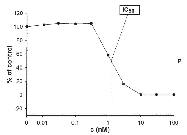
Figure 1. Graphic determination of IC50

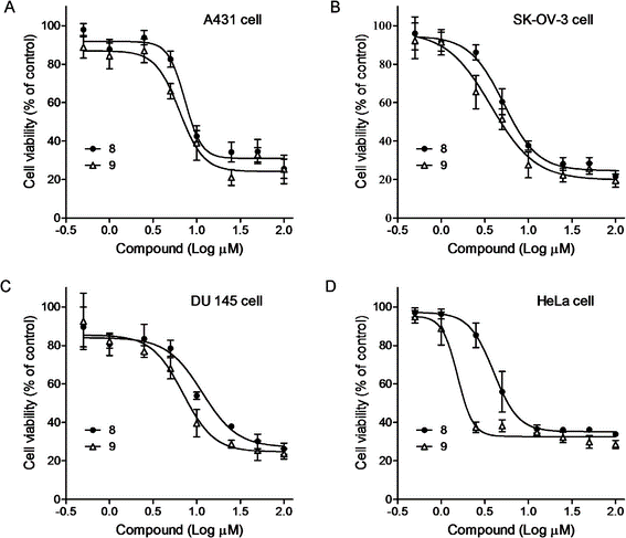
Figure 2. Effects of compounds on cell proliferation
Quote and Ordering
Please fill out the chart below or contact us to discuss your project with our experts and get a quote.
Payment methods supported: Invoice / Purchase Order, Wire Transfer, and Credit Card.
References
- Skehan, P., Storeng, R., Scudiero, D., Monks, A., McMahon, J., Vistica, D., Warren, J. T., Bokesch, H., Kenney, S., and Boyd, M. R. (1990) New colorimetric cytotoxicity assay for anticancer-drug screening. J. Natl. Cancer Inst. 82, 1107–1112.
- Perez, R. P., Godwin, A. K., Handel, L. M., and Hamilton, T. C. (1993) A comparison of clonogenic, microtetrazolium and sulforhodamine B assays for determination of cisplatin cytotoxicity in human ovarian carcinoma cell lines. Eur. J. Cancer 29A, 395–399.
- Griffon, G., Merlin, J. L., and Marchal, C. (1995) Comparison of sulforhodamine B, tetrazolium and clonogenic assays for in vitro radiosensitivity testing in human ovarian cell lines. Anticancer Drugs 6, 115–123.
- Papazisis, K. T., Geromichalos, G. D., Dimitriadis, K. A., and Kortsaris, A. H. (1997) Optimization of the sulforhodamine B colorimetric assay. J. Immunol. Methods 208, 151–158.
Explore Other Options
For research use only. Not for any other purpose.

