ONLINE INQUIRY
3T3-L1 Preadipocytes
Cat.No.: CSC-7692W
Species: Mouse
Source: Embryo
Cell Type: Preadipocyte
- Specification
- Background
- Scientific Data
- Q & A
- Customer Review
We now offer both Cryopreserved and Plated sub-confluent 3T3-L1 preadipocytes in 96 well format and media for proliferation, differentiation, maintenance of 3T3-L1 preadipocytes to adipocytes. We also offer media and reagent kits validated using 3T3-L1 and/or primary rodent preadipocytes and adipocytes. High quality control tested 3T3-L1 preadipocytes designed to work consistently using Bioarray's line of 3T3-L1 media.
The 3T3-L1 cell line is a continuous sub-strain of fibroblasts (preadipocytes) isolated from Swiss albino mouse embryos. In the state of undifferentiation, 3T3-L1 preadipocytes cells have a typical fibroblastic appearance. They were pike-shaped with long cytosol and many outgrowths, and the nucleus was oval and located at the center of the cell. When grown attached to the wall in a culture dish, these cells fuse to make a reticular pattern.
When activated by inducers (such as insulin, dexamethasone and 3-Isobutyl-1-methylxanthine (IBMX), cells can switch from fibroblast-like to adipocyte forms. This was the point at which they became very round and started collecting droplets of lipid, and as those droplets grew and fused, cell size increased exponentially, and now they morphed into a circular or oval adipocyte with many drops of lipid. And they facilitated both the formation and maintenance of lipid droplets, and the expression of lipid-metabolizing enzymes and transport proteins. Furthermore, 3T3-L1 preadipocytes are also an endocrine cell and release multiple adipocyte cytokines such as leptin, adiponectin, TNF-α, etc. Thus, 3T3-L1 cell lines are widely used to study the differentiation of adipocytes and their metabolism. They provide good laboratory models for metabolic disorders such as obesity and diabetes.
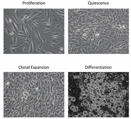 Fig. 1. Schematic representation of 3T3-L1 adipocyte differentiation (Ahmed M, Nguyen HQ, et al., 2018).
Ghrelin and IGF-1 Stimulated the Proliferation of Mouse 3T3-L1 Preadipocytes and Human Primary Preadipocytes
Obesity results mainly from the imbalance between energy production and expenditure that leads to adipose cells proliferating and growing. Ghrelin is a 28-amino-acid peptide that regulates energy expenditure, glucose and lipid metabolism. Phosphoinositol 3-kinase (PI3K)/Akt and extracellular regulated protein kinases (ERK)/mitogen-activated protein kinase (MAPK) regulate cell proliferation and differentiation via the insulin-like growth factor-1 (IGF-1) pathway. Ghrelin is known to enhance adipocyte growth and differentiation via MAPK/PI3K/Akt pathways, but it's not clear if this is accomplished by IGF-1.
Therefore, Miao's team examined the role of ghrelin in the proliferation and differentiation of mouse 3T3-L1 preadipocytes and human primary preadipocytes. Following 24 hours of incubation of 3T3-L1 preadipocytes with 0.01–1000 ng/ml ghrelin, 0.01 ng/ml ghrelin greatly increased cell proliferation and reached a maximum level at 10 ng/ml (Fig. 1A). For 100 and 1000 ng/ml, the effect dropped slightly but was still significantly greater than in the control group. To study the time course of ghrelin on 3T3-L1 preadipocyte proliferation, 3T3-L1 preadipocytes were treated with 10 ng/ml ghrelin for 4-48 hours. Between 4 to 48 hours, the OD value increased with 10 ng/ml ghrelin, peaking at 24 hours (Fig. 1B). Similar results were observed in human cells; a concentration of 1000 ng/ml ghrelin promoted human primary adipocyte proliferation after 24-hour treatment, effective from 24 to 48 hours, and subsiding by 72 hours (Fig. 1D). The CCK-8 and MTT assays confirmed these results. Similar to ghrelin administration, 0.25 ng/ml IGF-1 significantly promoted 3T3-L1 preadipocyte growth, peaking at 100 ng/ml (Fig. 1G). IGF-1 also demonstrated a similar concentration-dependent stimulatory effect in human cells (Fig. 1H).
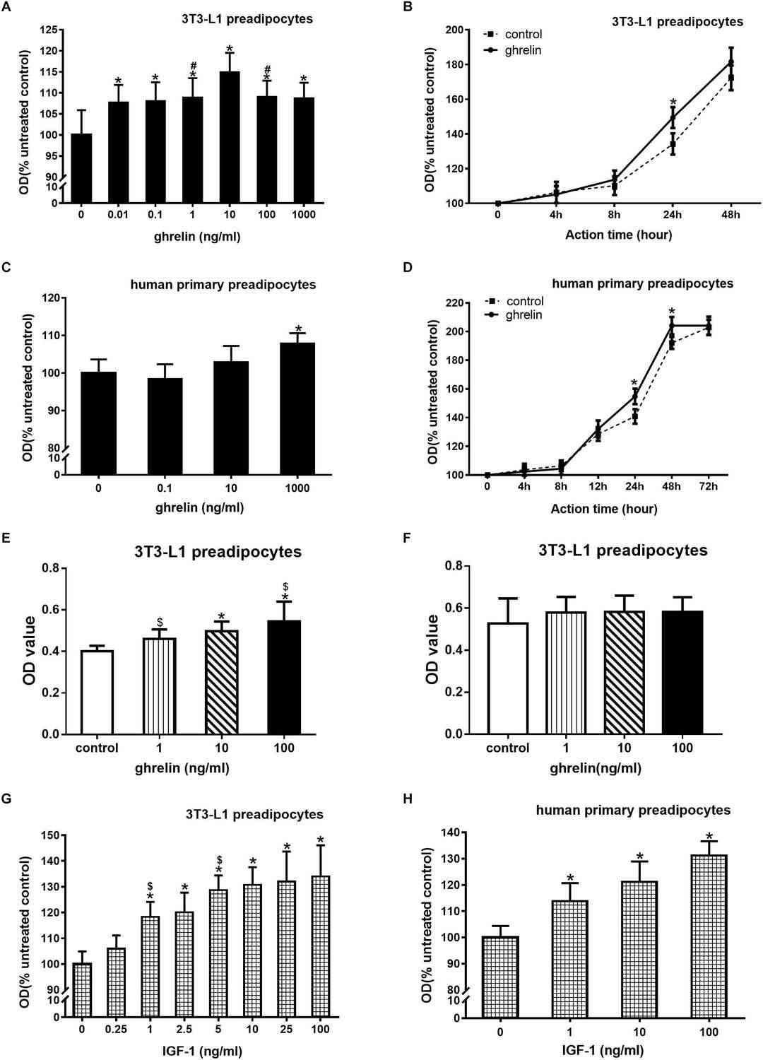 Fig. 1. Ghrelin and IGF-1 stimulated the proliferation of mouse 3T3-L1 preadipocytes and human primary preadipocytes (Miao H, Pan H, et al., 2019).
Overexpression of PVT1 Enhanced Adipogenesis in Preadipocytes
Obesity, a significant global health concern starting from childhood, is closely linked to the differentiation of preadipocytes and the process of adipogenesis. Long noncoding RNAs (lncRNAs), notably PVT1, play pivotal roles in regulating cellular functions. Although PVT1 is recognized for its oncogenic properties, its role in adipogenesis remains unclear. Zhang's team investigates the impact of lncRNA PVT1 on adipogenesis.
PVTI expression was first measured in various organs of mice, and the results showed a positive correlation between PVTI expression and organ fat content. In vitro, elevated PVTI expression coincided with the differentiation of mouse 3T3-L1 preadipocytes into mature adipocytes. To determine the association between PVT1 and the differentiation of 3T3-L1 preadipocyte, 3T3-L1 preadipocyte cell line with stable overexpression of PVT1 (LV-PVT1) and control cell line (LV-Vector) were acquired with the vector of lentivirus encoding PVT1 or with control lentivirus. Quantitative real-time PCR (qRT-PCR) revealed that PVT1 expression in successfully transfected 3T3-L1 preadipocytes was enhanced 50-fold (Fig. 2a). Overexpression of PVT1 was linked to increased TG accumulation (Fig. 2b). qRT-PCR results for adipocyte differentiation markers PPARγ, CEB/Pα, and AP2, measured on day 0 and day 6, showed upregulation in the LV-PVT1 group, consistent with previous findings (Fig. 2c–e). The CCK8 assay indicated PVT1 overexpression promoted 3T3-L1 preadipocyte proliferation (Fig. 2f). EdU staining showed more EdU positive cells in the LV-PVT1 group compared to control (Fig. 2g).
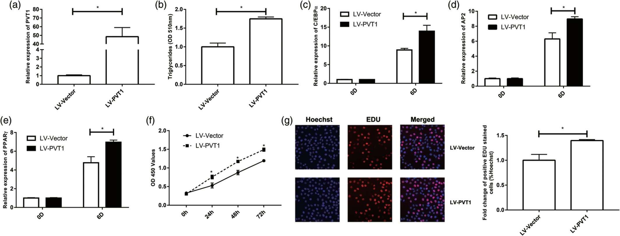 Fig. 2. The effect of PVT1 overexpression on adipogenesis. After 3T3-L1 preadipocyte was transfected with lentivirus encoding PVT1 (LV-PVT1) or negative control (LV-Vector) (Zhang L, Zhang D, et al., 2020).
In order to complete the differentiation process, you will need SuperCult® Preadipocyte Differentiation Medium Kit (cat# CM-1145X) and SuperCult® Adipocyte Medium (cat# CM-1230W), please order media according to your needs.
Oil droplets should appear within 4-7 days after differentiation is induced. They look extremely small initially, continue to accumulate and gradually fuse into several big locules.
No. At this time they are too sensitive to the stresses of shipping during differentiation. Only cryopreserved and sub-confluent preadipocytes are provided as live plated cells.
We provide 3T3-L1 preadipocytes in the following formats: 96-well, 48-well, 24-well, 8-chamber slides.
Use cells of a lower passage number. The 3T3-L1 cell line is NOT immortalized and is suitable until passage 12-13. Ensure cells are 100% confluent for 48 hours prior to initiating differentiation. Do not use fetal bovine serum during the proliferation process. It will affect later differentiation potential. We recommend using Creative Bioarray’s SuperCult® Preadipocyte Differentiation Medium Kit (cat# CM-1145X).
Ask a Question
Average Rating: 5.0 | 1 Scientist has reviewed this product
Successful subculture
The cell product I received was successfully subcultured.
13 May 2022
Ease of use
After sales services
Value for money
Write your own review

