ONLINE INQUIRY

Human Preadipocytes-subcutaneous
Cat.No.: CSC-7736W
Species: Human
Source: Adipose
Cell Type: Preadipocyte
- Specification
- Background
- Scientific Data
- Q & A
- Customer Review
Subcutaneous adipose tissue represents body fat situated between the skin and muscle layers and mainly consists of adipocytes while containing minor amounts of connective tissue and blood vessels and nerves. This tissue performs several essential functions in the human body which include temperature regulation while also protecting internal organs and acting as an energy reserve.
Human preadipocytes-subcutaneous originate from adult subcutaneous adipose tissue. These cells demonstrate polygonal or spindle-shaped morphology while possessing intercellular connections coupled with adhesiveness. When cultured in vitro preadipocytes demonstrate surface adherence and grow to establish either single or multiple layer cell colonies. Some preadipocytes start to mature into adipocytes over time by accumulating lipid droplets and acting as a reserve of adipocytes. Furthermore, preadipocytes have the capability to produce different bioactive compounds like cytokines and hormones which help in maintaining metabolic and immune system functions throughout the body. These factors show a strong link to the development and advancement of diseases like obesity, diabetes and cardiovascular conditions. Therefore, researchers use this cell line to study both adipocyte differentiation mechanisms and lipid metabolism together with adipose tissue's ecological features. And this cell line functions as a screening platform for potential drugs targeting obesity, diabetes, and cardiovascular conditions.
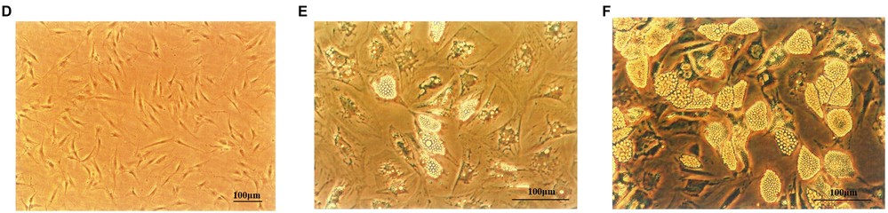 Fig. 1. Human primary preadipocytes proliferation and differentiation. (D) Human primary preadipocyte on Day 8 (×100). Adipocytes on Day 10 (E) and Day 18 (F) after inducing differentiation (×200) (Miao H, Pan H, et al., 2019).
Hypoxanthine Secretion from Human Adipocytes
Hyperuricemia, associated with obesity and metabolic disorders, involves a delicate balance in purine metabolism. Recent murine studies reveal that adipose tissue contributes to uric acid production. Nagao et al. investigated human adipocytes and human adipose tissue by culturing freshly isolated hWAT and mWAT and measuring secreted metabolite levels. Analyzing gene expressions related to purine metabolism and metabolites under hypoxia in adipocytes further refines this understanding.
They assessed purine metabolite levels in cultured media from differentiated adipocytes via HPLC-UV. Human preadipocytes-subcutaneous were differentiated as per the manufacturer's protocol. Oil red O staining shows human preadipocytes (day 0) and differentiated adipocytes (day 8) in Supporting Information Figure 2A. Uric acid and xanthine were undetectable, but hypoxanthine was present and increased during differentiation (Fig. 1A, Fig. 2B). Given that obese adipose tissues are often hypoxic, we explored hypoxia's effect on human adipocytes. Under 1% O2 for 24 hours, hypoxanthine levels significantly rose compared to normoxia (Fig. 1A), suggesting enhanced de novo purine synthesis under hypoxia. In murine 3T3-L1 adipocytes, uric acid, not hypoxanthine or xanthine, increased in hypoxia (Fig. 1B), a pattern observed in primary murine adipocytes from subcutaneous WAT (Fig. 2C).
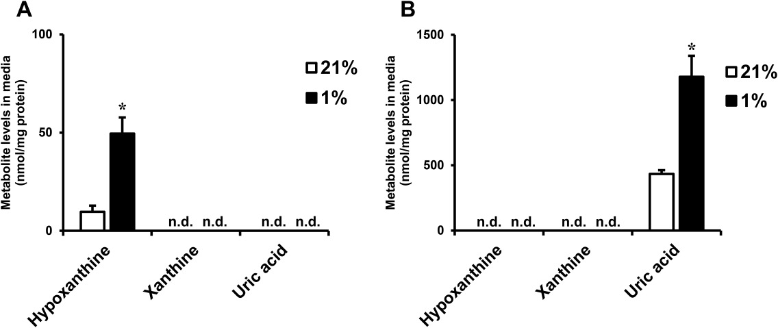 Fig. 1. Hypoxanthine levels in culture media from human adipocytes (Nagao H, Nishizawa H, et al., 2018).
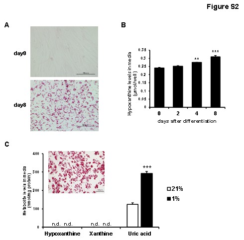 Fig. 2. Hypoxanthine levels in culture media from human preadipocytes and differentiated adipocytes (Nagao H, Nishizawa H, et al., 2018).
Ghrelin and IGF-1 Stimulated the Proliferation of Mouse 3T3-L1 Preadipocytes and Human Primary Preadipocytes
Obesity is tied to adipocyte proliferation influenced by transcription factors and hormones. Ghrelin, a known appetite regulator, affects adipocyte function, yet its mechanisms are not fully understood. Miao's team investigated ghrelin's role in 3T3-L1 and human preadipocyte proliferation and differentiation using MTT assay, Oil Red O staining, RT-PCR, and glycerol-3-phosphate dehydrogenase assay.
3T3-L1 preadipocytes were treated with ghrelin (0.01–1000 ng/ml) for 24 hours. Figure 3A shows that 0.01 ng/ml ghrelin significantly enhanced cell growth (107.7 ± 4.2 vs. 100.0 ± 5.9, p < 0.05), with maximal effect at 10 ng/ml (114.9% of control, p < 0.05). Higher concentrations (100 and 1000 ng/ml) slightly reduced the effect, but it remained significant (p < 0.05). Similarly, ghrelin increased proliferation in human primary preadipocytes, as shown in Figure 3C. Only 1000 ng/ml significantly boosted growth (107.8 ± 2.8 vs. 100.0 ± 3.6, p < 0.05), with no effect at lower doses (0.1 and 10 ng/ml). Figure 3D illustrates the OD value increase over 72 hours with 1000 ng/ml ghrelin, peaking at 48 hours (204.1 ± 6.1 vs. 192.1 ± 4.2, p < 0.05), disappearing by 72 hours.
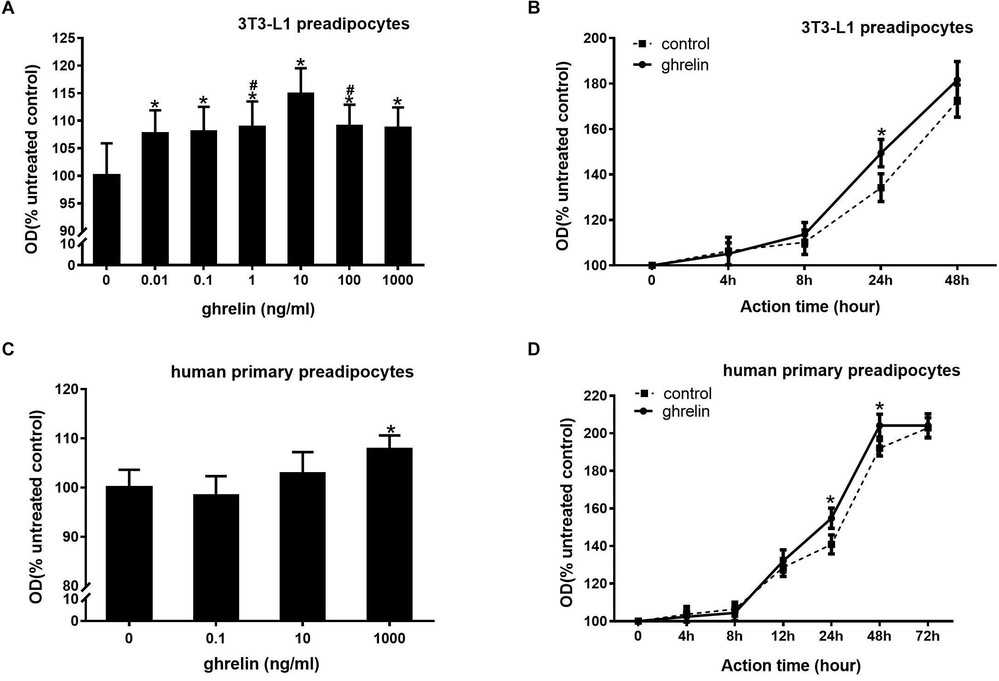 Fig. 3. Ghrelin and IGF-1 stimulated the proliferation of mouse 3T3-L1 preadipocytes and human primary preadipocytes (Miao H, Pan H, et al., 2019).
The preadipocytes are isolated from human subcutaneous adipose tissue. The adipocytes are differentiated in vitro using these isolated preadipocytes.
Preadipocytes can be trypsinized and replated several times. Preadipocytes grow slower with each passage and differentiate poorly after passage 4. Cells are shipped at Passage 2-4.
You can order preadipocytes and pre-made culture media kit (cat# CM-1145X) for adipocyte differentiation. The protocol for differentiating the cells can be found in our manual.
Yes, we often have cells from various depots available. Please inquire as to price and availability.
Ask a Question
Average Rating: 5.0 | 1 Scientist has reviewed this product
Easy recovering
During our experiments, the cell product was easily recovered.
19 May 2022
Ease of use
After sales services
Value for money
Write your own review

Description: Preadipocytes are isolated from omental, mesenteric, or perirenal adipose tissue and are tested for their ability to differentiate in culture to mature adipocytes. Preadipocytes are available cryopreserved or plated and mature adipocytes are available in a variety of plated formats.
The greater omentum is the largest peritoneal fold within the abdomen and has an immunologic function. The omentum aids in isolating peritoneal infection and absorbing contaminants through the mesothelial stomata. Adipokines and fatty acids from this depot have direct access to the liver through the portal circulation which may lead to hepatic dysfunction.

Description: Preadipocytes are isolated from omental, mesenteric, or perirenal adipose tissue and are tested for their ability to differentiate in culture to mature adipocytes. Preadipocytes are available cryopreserved or plated and mature adipocytes are available in a variety of plated formats.
Perirenal adipose resides near the kidneys outside the peritoneal cavity. Fat from this depot has not been correlated with increased risk for metabolic disease.

Description: Visceral preadipocytes can be cultured as growing precursor cells or differentiated into adipocytes using medium supplemented with adipogenic and lipogenic hormones. This instruction manual describes procedures required to induce human preadipocytes to differentiate into mature adipocytes as well as culturing methods for human preadipocytes and adipocytes
Description: 3T3-L1 murine adipocytes have been fundamental in metabolic disease research for over 30 years. Originally derived from Swiss mouse embryo tissue by Dr. Howard Green of Harvard Medical School, the 3T3-L1 system has been pivotal in advancing the understanding of basic cellular mechanisms associated with diabetes, obesity and related disorders.
We now offer both Cryopreserved and Plated sub-confluent 3T3-L1 preadipocytes in 96 well format and media for proliferation, differentiation, maintenance of 3T3-L1 preadipocytes to adipocytes. We also offer media and reagent kits validated using 3T3-L1 and/or primary rodent preadipocytes and adipocytes. High quality control tested 3T3-L1 preadipocytes designed to work consistently using Bioarray's line of 3T3-L1 media.
Description: Microvascular endothelial cells line blood vessels and contribute to many biological processes such as angiogenesis, coagulation, trafficking of lymphocytes, and the inflammatory response. Microvascular endothelial cells are diverse and have specific cellular characteristics and functions depending on the organ/tissue in which they are found. Adipose tissue is unique because it has the capacity to continually grow throughout adult life. It thus has a high occurrence of angiogenesis in order to provide the extensive vascularization require for adipose tissue. Studies have shown that angiogenesis precedes adipogenesis, implying that microvascular endothelial cells influence the proliferation of preadipocytes. Microvascular endothelial cell growth is at the same time stimulated by adipocyte secreted VEGG, suggesting a complex paracrine relationships between microvascular endothelial cells and preadipocytes during tissue development.HAMEC from Bioarray Research Laboratories are isolated from human adipose tissue. HAMEC are cryopreserved after purification and delivered frozen. Each vial contains >1 x 10^6 cells in 1 ml volume. HAMEC are characterized by immunofluorescent method with antibodies to vWF/Factor VIII and CD31 (P-CAM) and by uptake of DiI-Ac-LDL. HAMEC are negative for HIV-1, HBV, HCV, mycoplasma, bacteria, yeast and fungi. HAMEC are guaranteed to further expand for 15 population doublings at the conditions provided by Bioarray Research Laboratories.
Description: Preadipocytes, found in adipose tissues throughout adulthood, can proliferate and differentiate into mature adipocytes based on energy balance, which in turn increases adipose tissue mass. In vitro studies show that preadipocytes from different tissues vary in lipid accumulation, expression of adipogenic transcription factors, and susceptibility to TNF-induced apoptosis. Furthermore, adipocyte differentiation is closely linked to various physiological and pathological processes, including fat metabolism, energy balance, obesity, diabetes, hyperlipidemia, and breast cancer.
Human Preadipocytes - Visceral (HPAs-v) from Cretive Bioarray are isolated from human visceral fat tissue. HPA-v are cryopreserved at passage one and delivered frozen. Each vial contains at least 0.5 x 10^6 cells in 1 ml volume. HPAs-v are negative for HIV-1, HBV, HCV, mycoplasma, bacteria, yeast and fungi. HPAs-v are guaranteed for further culture under conditions specified by Creative Bioarray.
