Featured Products
Our Promise to You
Guaranteed product quality, expert customer support

ONLINE INQUIRY
CAL-33
Cat.No.: CSC-C0479
Species: Human
Source: tongue squamous cell carcinoma
Morphology: epitheloid cells growing adherent in monolayers
Culture Properties: monolayer
- Specification
- Background
- Scientific Data
- Publications
- Q & A
- Customer Review
Immunology: cytokeratin +, cytokeratin-7 -, cytokeratin-8 +, cytokeratin-17 +, cytokeratin-18 +, cytokeratin-19 -, desmin -, endothel -, EpCAM -, GFAP -, neurofilament -,
The CAL-33 cell line is an adherent cell line derived from human tongue squamous carcinoma tissue. This cell line was originally established in 1983 from a moderately differentiated squamous cell tongue cancer in a 69-year-old male patient, obtained from surgically resected fragments of tongue damage. CAL-33 cells exhibit typical epithelioid cell-like morphology and grow as a monolayer adherent to the wall in culture, with remarkable proliferative capacity and the ability to divide rapidly under suitable culture conditions, allowing for rapid growth in number.
This cell line exhibits strong invasiveness and is capable of secreting a variety of proteases, such as matrix metalloproteinases. These enzymes create conditions for cell migration and invasion by degrading the extracellular matrix. In the Cancer Genome Atlas (CCLE), the CAL-33 cell line is listed as a cell line with potential for genomic and protein analysis under reference number 18232. In addition, CAL-33 cells exhibit tumorigenicity in nude mice. Due to these properties, CAL-33 cells are widely used to study the mechanisms of tongue cancer development and progression, to assess the efficacy of new drugs on tongue cancer cells, and to analyze the expression and function of cancer-related genes. This cell line has also been used to study radiosensitivity and tolerance and as a model for head and neck squamous cell carcinoma studies.
 Fig. 1. Representative images of Cal33 cells (Emer C, Hildebrand LS, et al., 2023).
Grape Cane Extract Inhibits Cell Viability of HNSCC Cells
Plants are abundant sources of biologically active molecules, with phenolic compounds notably recognized for their antioxidant and anticancer properties. Despite their potential health benefits, grape canes - the by-product of vine pruning - have been underutilized, although they contain valuable phenolic antioxidants. Head and neck squamous cell carcinoma (HNSCC) is a common cancer with resistance to conventional treatments. Polyphenols, known for their anticancer potential and low toxicity, offer promising alternatives. Therefore, Squillaci et al. evaluated the antioxidant and anticancer efficacy of phenolic extracts from "Greco" grape canes on HNSCC cells. First, the antiproliferative activity of "Greco" grape cane extracts on oral Cal-33 and laryngeal JHU-SCC-011 HNSCC cell lines was assessed using an MTT assay. Treatment with extracts prepared at pH 7 for 60 minutes led to a marked dose- and time-dependent inhibition of cell proliferation, achieving an IC50 of 0.25 mg/mL after 72 hours (Fig. 1). Further analysis identified that the polyphenol-rich fraction FR 5 exhibited significant cytotoxicity, with IC50 values of 0.10 mg/mL and 0.12 mg/mL for Cal-33 and JHU-SCC-011 cells, respectively, approximately half the concentration needed by the crude extract for similar effects (Fig. 2). This indicates that polyphenols are key contributors to the antiproliferative effects of the extracts.
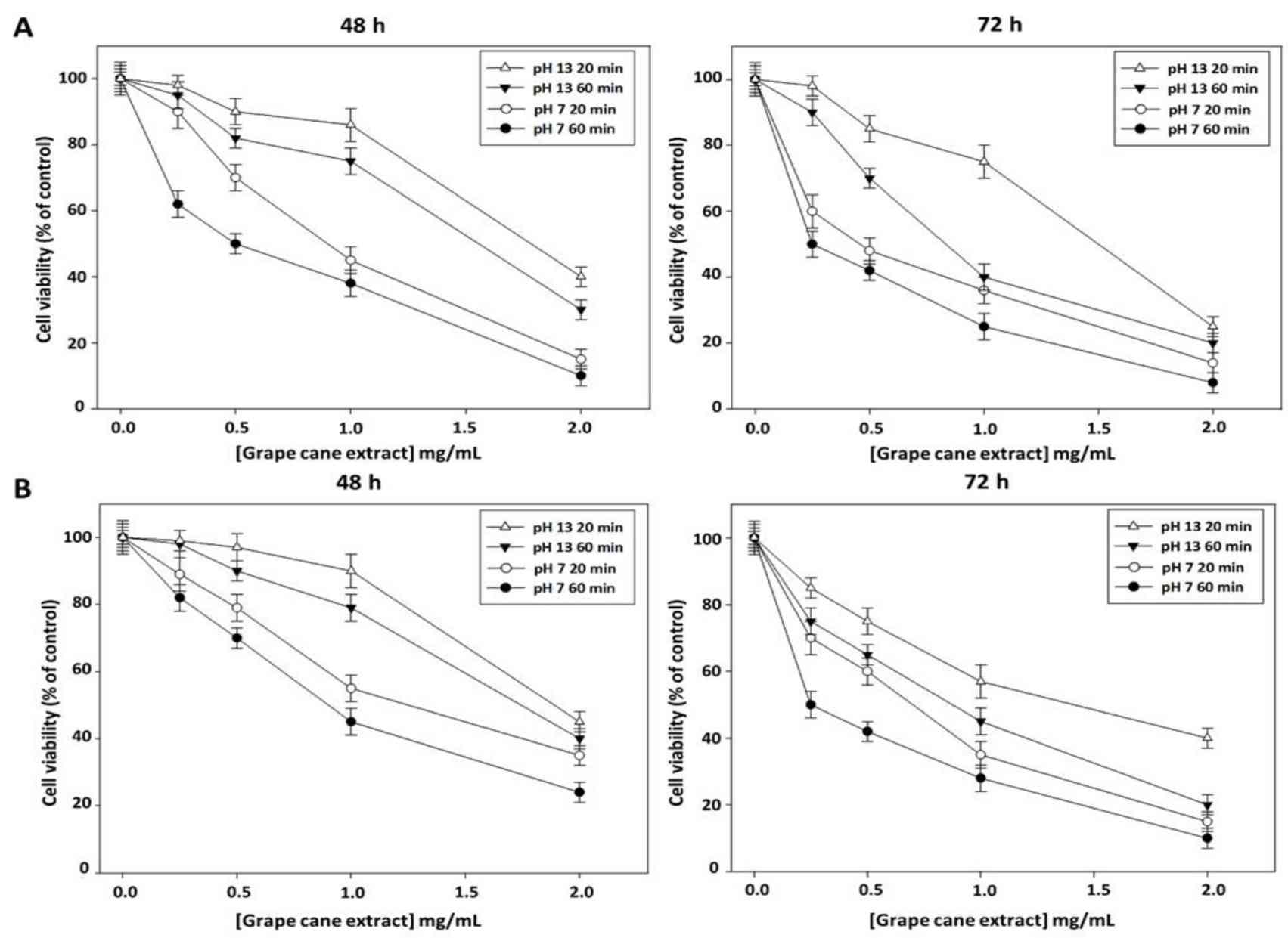 Fig. 1. Effect of "Greco" grape cane extracts prepared at different pHs and extraction times on cell viability of Cal-33 and JHU-SCC-011 cell lines (Squillaci G, Vitiello F, et al., 2022).
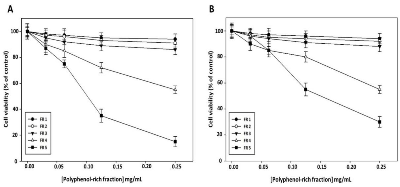 Fig. 2. Effect of polyphenol-rich fractions on HNSCC cell viability (Squillaci G, Vitiello F, et al., 2022).
Apoptosis Assessment in Cal-33 and JHU-SCC-011 Cell Lines Treated with AdoMet
S-Adenosyl-l-methionine (AdoMet) is a multifunctional biomolecule, involved in many biological processes, especially as a main methyl donor. It has antiproliferative and proapoptotic effects on multiple types of cancers, and recent studies have shown its utility for gastric, prostate and head and neck squamous tumours (HNSC). But the exact molecular basis of AdoMet's activity and its influence on HNSC remains unknown. Mosca et al. tested the apoptotic effect of AdoMet on Cal-33 and JHU-SCC-011 cell lines. Double-labeled with Annexin V-FITC and PI, FACS analysis determined the degree of cell death. The findings indicated that, in Cal-33 cells, at least a quarter of the cells had become dead after 48 hours (Fig. 3a), while after 72 hours of infection, approximately 19% of JHU-SCC-011 cells were dead (Fig. 3b). The AdoMet treatment knocked down full-length caspase 9 (cause of apoptosis), caspase 6, and caspase 7 (upstream caspases) in both cell lines, as well as cleavage of PARP, with an especially dramatic effect at 72 hours (Fig. 4). Both lines of cells had higher levels of proapoptotic Bax and lower levels of antiapoptotic Bcl-2 when treated with AdoMet. This shift in the ratio of Bax to Bcl-2 points towards a caspase-dependent apoptotic response. Finally, AdoMet promotes apoptosis in Cal-33 and JHU-SCC-011 by a caspase-dependent pathway with a higher Bax/Bcl-2 ratio (Fig. 4).
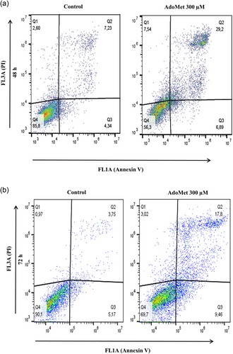 Fig. 3. Representative dot plots of Annexin V-FITC and PI-stained head and neck cancer cells (Mosca L, Pagano M, et al., 2019).
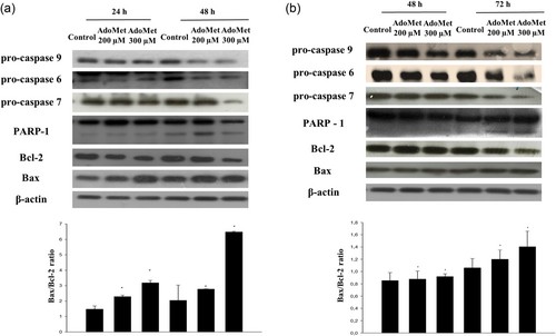 Fig. 4. Effects of AdoMet on the expression levels of relevant proteins regulating apoptotic pathways in head and neck cancer cells (Mosca L, Pagano M, et al., 2019).
Mouth cancers most commonly begin in the flat, thin cells (squamous cells) that line your lips and the inside of your mouth. Most oral cancers are squamous cell carcinomas.
Ask a Question
Average Rating: 5.0 | 1 Scientist has reviewed this product
Cost-effective
We have found the tumor cell products at Creative Bioarray to be highly cost-effective.
12 Aug 2023
Ease of use
After sales services
Value for money
Write your own review
- You May Also Need

