Featured Products
Our Promise to You
Guaranteed product quality, expert customer support

ONLINE INQUIRY
TF-1
Cat.No.: CSC-C0395
Species: Homo sapiens ,human
Source: bone marrow
Morphology: lymphoblast
- Specification
- Background
- Scientific Data
- Q & A
- Customer Review
CSF1PO:13,13
D13S317: 8,9
D16S539:9,12
D5S818:13
D7S820:12
TH01:7,9
TPOX:8
vWA:15,17
D3S1358: 15
D21S11: 30
D18S51: 13
Penta E: 5,17
Penta D: 10,13
D8S1179: 11,15
FGA: 18,19
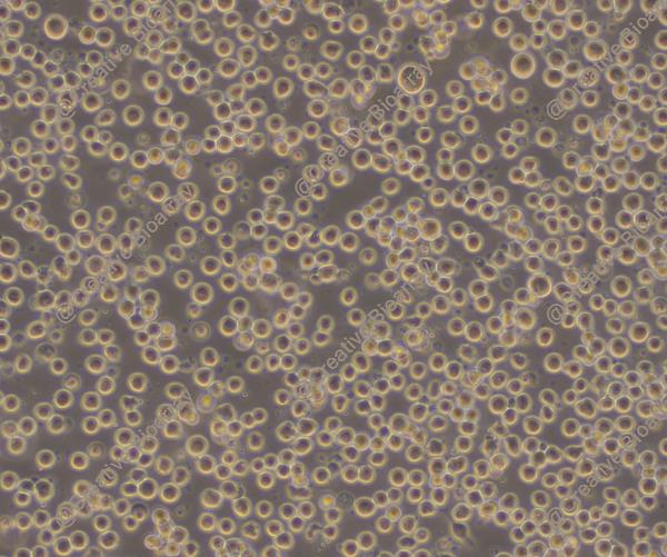
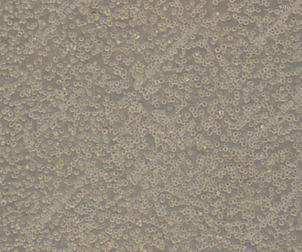
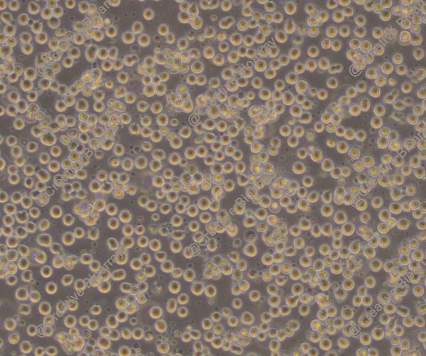
The TF-1 cell line was generated from erythroblast cells extracted from the bone marrow of a 35-year-old Asian man with severe pancytopenia in 1987. In their morphology, TF-1 cells are lymphoblast-like cells. And they don't produce glycophorin A or carbonic anhydrase I. These cells are obviously erythroid cells, according to their cell shape, cytochemistry and constitutive globin gene expression. Hemin and δ-aminolevulinic acid can also activate TF-1 cells to create hemoglobin and TPA (12-O-Tetradecanoylphorbol-13-acetate) can really help the cells to become macrophages. In vitro, TF-1 cells are totally dependent on IL-3 (interleukin 3) or GM-CSF (granulocyte-macrophage colony-stimulating factor) for growth and respond no to IL-5. They typically exhibit semi-adherent and semi-suspension growth characteristics, meaning that some cells adhere to the culture flask wall while others remain suspended in the medium. That makes the cell line relatively straightforward to cultivate.
What makes the TF-1 cell line unique is that it is sensitive to hundreds of different cytokines including IL-1, IL-4, IL-6, IL-9, IL-11, IL-13, stem cell factor (SCF), leukemia inhibitory factor (LIF) and nerve growth factor (NGF). This responsiveness creates a wonderful model system for studying myeloid progenitor-cell proliferation and differentiation. Hence, TF-1 cell line has a huge usage in 3D cell culture, immunological disorder and immunology research. TF-1 cells are also used to test drug effects on leukemic cells, which helps screen and evaluate treatments against hematological cancers such as leukemia. This is why the TF-1 cell line becomes an essential component in the production of new drugs.
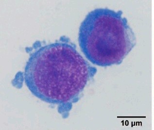 Fig. 1. Wright–Giemsa-stained TF-1 cells (Peng, Y., Lu, C., et al., 2021).
CP-17 Inhibits the Proliferation and Restores the EPO-Induced Differentiation in TF-1(IDH2/R140Q) Cells
The IDH2/R140Q mutation contributes to acute myeloid leukemia (AML) by accumulation of oncometabolite D-2-HG, making IDH2 a promising target for AML treatment. Chen et al. discovered and studied a novel IDH2/R140Q inhibitor (CP-17), which effectively inhibits intracellular D-2-HG production and suppresses the proliferation of IDH2/R140Q mutant cells. Experimental evidences are as follows: TF-1(IDH2/R140Q) cells can grow in the absence of GM-CSF, while the wild-type TF-1 [TF-1(WT)] cell growth was GM-CSF-dependent. Cellular proliferation assay suggested that treatment with 3 μM CP-17 significantly inhibited the GM-CSF-independent growth of TF-1(IDH2/R140Q) cells but had no obvious effect on the proliferation of TF-1(WT) cells (Fig. 1). Similar to human tumor cells, TF-1 (IDH2/R140Q) cells produce high levels of D-2-HG. CP-17 exhibited dose-dependent inhibition of intracellular D-2-HG in TF-1 (IDH2/R140Q) cells (Fig. 2a). EPO-induced differentiation in TF-1 (WT) cells was marked by a red color change and increased hemoglobin γ expression (Fig. 2b). This differentiation was absent in TF-1 (IDH2/R140Q) cells, due to high D-2-HG levels, indicating that the IDH2/R140Q mutation blocked EPO-induced differentiation. Treatment with 1 μM CP-17 reduced intracellular D-2-HG levels and restored the EPO-induced color change and hemoglobin γ expression in TF-1 (IDH2/R140Q) cells, suggesting restored differentiation.
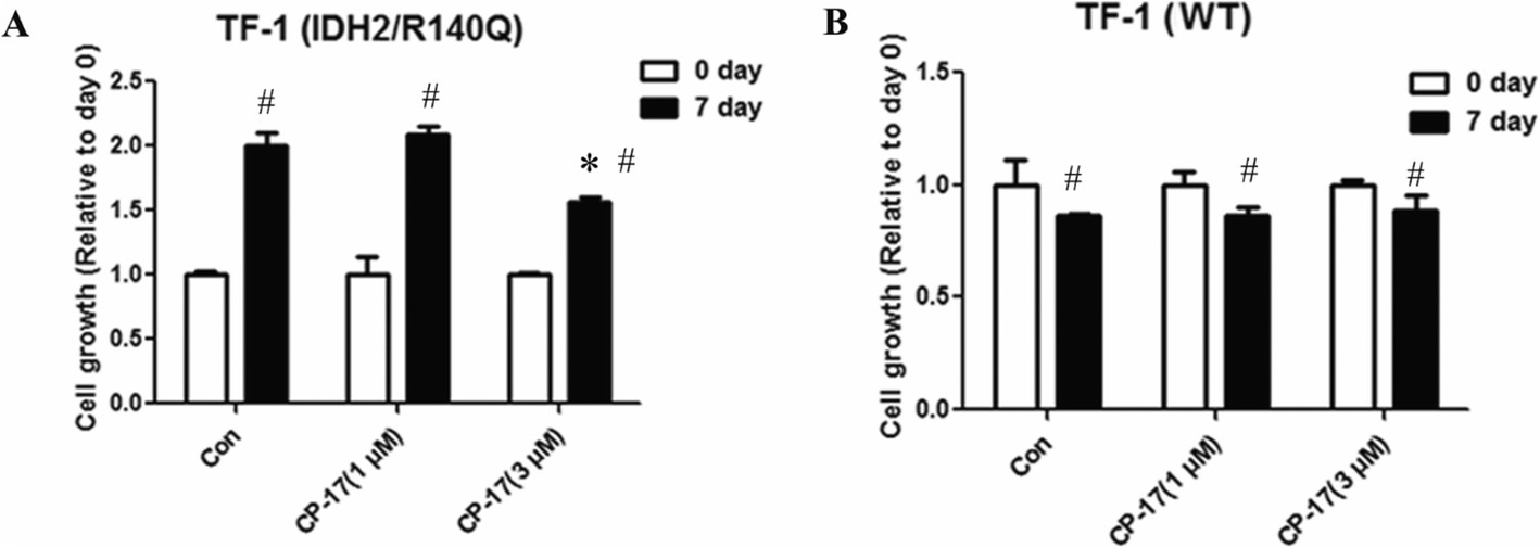 Fig. 1. CP-17 treatment reversed GM-CSF-independent growth induced by IDH2/R140Q. IDH2/R140Q expression confers GM-CSF independent growth of the TF-1 cells (Chen J., Yang J., et al., 2020).
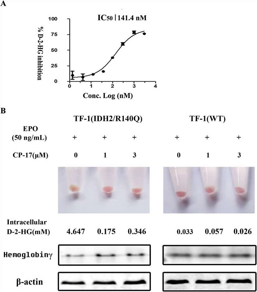 Fig. 2. CP-17 reversed the TF-1(IDH2/R140Q) differentiation block (Chen J., Yang J., et al., 2020).
CCRL2 Overexpression Increases the Resistance to MDS-L and TF-1 Cells to Azacitidine
DNMT inhibitors are the standard treatment for high-risk myelodysplastic syndrome (MDS) and secondary acute myeloid leukemia (sAML), but patient survival remains low, and resistance mechanisms are unclear. Karantanos et al. found that CCRL2 (an atypical chemokine receptor) overexpression in MDS/sAML cell is linked to poor response to DNA methyltransferase (DNMT) inhibitors, and its knockdown enhances the efficacy of azacitidine. The effect of CCRL2 expression on the development of azacitidine resistance was analyzed by introducing pLV-Puro-TRE-CCRL2 and pLV-HygroCMV>tTS/rtTA lentiviruses into MDS-L cells. Doxycycline-induced CCRL2 expression was confirmed through western blot and flow cytometry (Fig. 3A). CCRL2 overexpression reduced the clonogenicity inhibition by 0.5 mM azacitidine and significantly raised azacitidine's IC50 value (Fig. 3B). It also slightly lessened the apoptotic effect of 0.5 and 1 mM azacitidine (Fig. 3C). Using CRISPR-Cas9 via electroporation, CCRL2 was deleted in TF-1 cells. Clonogenicity assays showed that CCRL2 deletion reduced TF-1 cell clonogenicity. TF-1 cells with CCRL2 deletion were again transduced with the same lentiviruses. Doxycycline-induced CCRL2 expression was verified (Fig. 3D), showing that CCRL2 overexpression decreased clonogenicity inhibition by 0.5 and 1 mM azacitidine and increased the IC50 value (Fig. 3E).
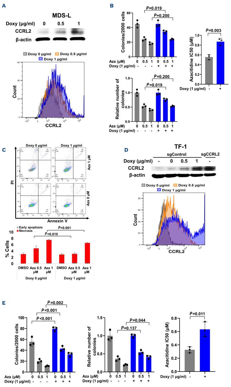 Fig. 3. Doxycycline-induced CCRL2 overexpression increases the resistance of MDS-L and TF-1 cells to azacitidine (Karantanos T., Teodorescu P., et al., 2023).
Permeable nucleic acid stains that can penetrate cell membranes can be used to stain both living and fixed cells without the need for permeabilization. On the other hand, impermeable dyes selectively stain dead cells with disrupted membranes, but not viable cells.
Ask a Question
Average Rating: 5.0 | 1 Scientist has reviewed this product
Reliable
Creative Bioarray's products have proven to be reliable and consistent in scientific research.
07 Nov 2022
Ease of use
After sales services
Value for money
Write your own review
- You May Also Need

