Featured Products
Our Promise to You
Guaranteed product quality, expert customer support

ONLINE INQUIRY
HCT-116
Cat.No.: CSC-C0586
Species: Human
Source: colon carcinoma
Morphology: epithelial-like small cells growing as monolayer
Culture Properties: monolayer
- Specification
- Background
- Scientific Data
- Publications
- Q & A
- Customer Review
Immunology: cytokeratin +, cytokeratin-7 +, cytokeratin-8 +, cytokeratin-17 -, cytokeratin-18 +, cytokeratin-19 +, desmin -, endothel -, EpCAM +, GFAP -, neurofilament -, vimentin -
Viruses: PCR:
The HCT-116 cell line is a widely used model system in cancer research, particularly for the study of colorectal cancer. This cell line was established from the primary colon carcinoma of an adult male patient, making it a clinically relevant model for investigating the molecular mechanisms and therapeutic vulnerabilities of this common type of gastrointestinal malignancy.
A distinguishing feature of the HCT-116 cell line is the presence of a mutation in the RAS oncogene, specifically at codon 13. The RAS family of genes, including KRAS, NRAS, and HRAS, play crucial roles in regulating cell growth, proliferation, and survival. Mutations in these genes, such as the one found in HCT-116 cells, can lead to the constitutive activation of the RAS signaling pathway, which is a hallmark of many types of cancer, including colorectal cancer.
The tumorigenic potential of the HCT-116 cell line has been well-established through studies demonstrating its ability to form tumors when transplanted into nude mice. This characteristic makes the HCT-116 model a valuable tool for investigating the in vivo behavior of colorectal cancer cells, including their growth dynamics, metastatic potential, and response to various therapeutic interventions.
Over the years, the HCT-116 cell line has become a widely recognized and extensively studied model in the field of colorectal cancer research. Researchers have utilized this cell line to elucidate the complex molecular pathways and genetic alterations that drive the development and progression of this type of cancer. The availability of the HCT-116 model has been instrumental in advancing our understanding of the fundamental biology of colorectal tumors and has contributed to the identification of potential therapeutic targets and the development of novel treatment strategies.
Recombinant Chemokines and Supernatants Collected From HCT-116 Colorectal Cancer Cells Stimulate the Mobilization of Intracellular Ca2+ in NK92 Cells
It is known that there is a strong association between chemotaxis and the mobilization of intracellular calcium. To confirm such principles in NK92 cells, the mobilization of intracellular Ca2+ was assessed. First, Triton-X was used as a control to induce the release of intracellular calcium and EGTA to chelate this response. Next, two different concentrations of CXCL16, CCL27, and CCL28 were examined, namely 10 and 100 ng/mL, and observed that the 100 ng/mL concentration of the three chemokines induced the mobilization of (Ca2+); however, only the 10 ng/mL concentration of CCL28 produced a positive response. Intriguingly, the undiluted, 1:1 dilution or 1:10 dilution of supernatants collected after 24 or 48 h from HCT-116 cells also provoked the mobilization of (Ca2+) in NK92 cells.
To verify whether the supernatants collected from HCT-116 may utilize similar receptors to that of recombinant chemokines, desensitization experiments were performed. To gain insight into this issue, fura-2-AM-labeled NK92 cells were incubated with undiluted supernatants collected from HCT-116 cells after 24 h or 48 h incubation. The results indicate that addition of 24 h supernatants entirely desensitized the effects of the 24 h supernatants, the CXCL16 and CCL28, but there were partial desensitization of the 48 h supernatants and no effect on CCL27 (Fig. 1a). However, addition of the 48 h supernatants desensitized the calcium mobilization effects of the 48 h supernatants, 24 h supernatants, CXCL16, CCL27, and CCL28 (Fig. 1a). The reciprocal experiments were performed where recombinant chemokines were added before the addition of the supernatants. CXCL16 desensitized CXCL16 activity but not the activity of the supernatants (Fig. 1b). Similarly, CCL27 and CCL28 desensitized CCL27 and CCL28 activity, respectively, but not the supernatants with the exception where CCL28 partly desensitized the effect of 24 h supernatants (Fig. 1b). These results confirm that 24 h supernatants contain CXCL16 and CCL28, whereas 48 h supernatants contain all the three chemokines examined.
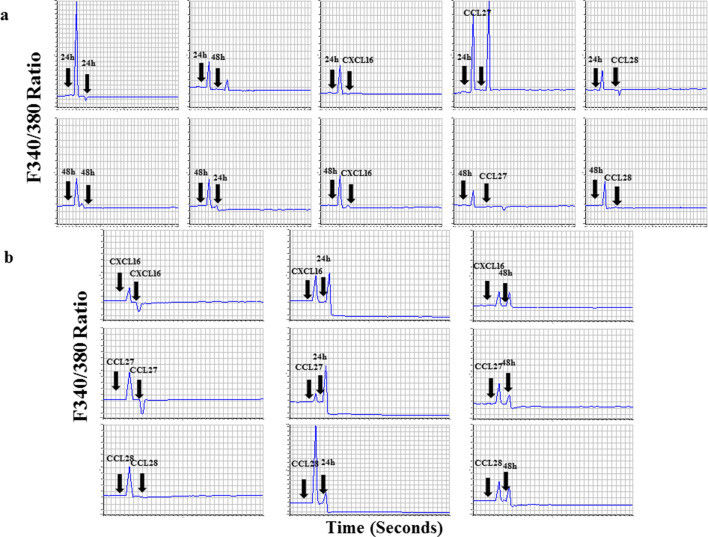 Fig. 1 Desensitization of chemokine receptors with recombinant chemokines and HCT-116 supernatants. (Elemam NM, et al., 2019)
Overexpression of MDR1 Induces Drug Resistance Phenotype in HCT-116 Cells
The MDR1 gene was transfected into HCT-116 cells and analyzed the resulting transfected cells (HCT-116/R) for the drug-resistant phenotype under a microscope. The cells overexpressing MDR1 had more intracellular vacuoles compared to the wild-type cells (HCT-116) (Fig. 2a). Next, the ability of HCT-116/R cells to reduce the drug uptake was analyzed using rhodamine 123 staining. Most of the HCT-116 cells showed intense rhodamine 123 fluorescence, whereas the fluorescence of HCT-116/R cells was weaker, which indicates that the uptake of rhodamine 123 by the MDR1-overexpressing cells was reduced (Fig. 2b). The intracellular distribution and accumulation of doxorubicin were analyzed in both cell lines and found that most of HCT-116 cells displayed prominent doxorubicin fluorescence in the nucleus, while HCT-116/R cells showed no doxorubicin fluorescence in the nucleus and diminished fluorescence in the cytoplasm (Fig. 2c). The overexpression of MDR1 in HCT-116/R cells was also confirmed by immunoblotting (Fig. 2d). Moreover, expression of HIF-1α and Nrf-2 was also upregulated. These results suggest that the elevated expression of MDR1, HIF-1α, and Nrf-2 is associated with the development of drug resistance.
The chemosensitivity of HCT-116 and HCT-116/R cells was determined to 5-FU. The half-maximal inhibitory concentration (IC50) of 5-FU for HCT-116 and HCT-116/R was ~241.40 and ~702.30 µM, respectively (Fig. 2e), which indicates that the HCT-116/R cells are highly resistant to 5-FU. Therefore, the MDR1 overexpression confers drug resistance in human colorectal carcinoma (HCT-116) cells.
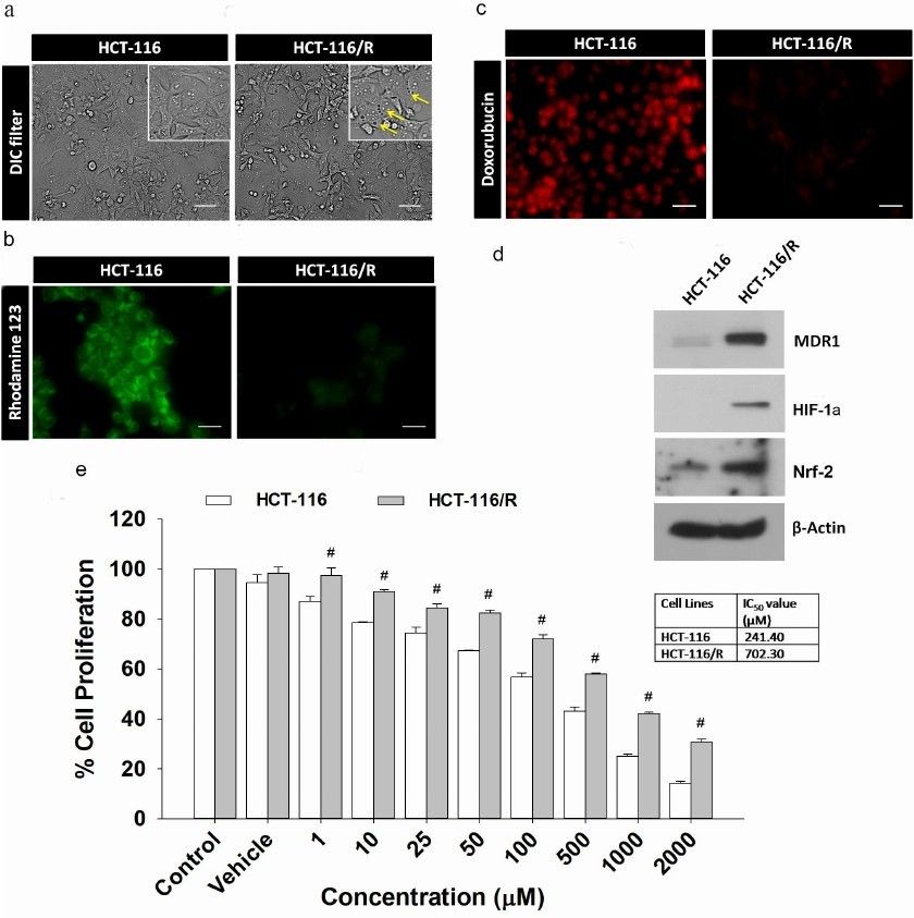 Fig. 2 Characterization of drug-resistant HCT-116/R cells obtained by transfection of HCT-116 cells with the MDR1 gene. (Waghela BN, et al., 2021)
Oncogenes are normal genes that delay cell division, repair DNA errors, or tell cells when to die (i.e., apoptosis or programmed cell death).
The HCT-116 cell line was derived from the primary colon carcinoma of an adult male patient.
HCT-116 cells carry a mutation in the KRAS oncogene at codon 13, which leads to the constitutive activation of the RAS signaling pathway.
HCT-116 cells are highly tumorigenic and can form tumors when transplanted into nude mice, making them a valuable model for studying the in vivo characteristics of colorectal cancer.
Ask a Question
Average Rating: 4.7 | 3 Scientist has reviewed this product
Easier experiments
This product has made my experiments easier!
24 Jan 2022
Ease of use
After sales services
Value for money
Exceptional quality
The cells I've received have always been of exceptional quality, with consistent growth rates and maintenance of the KRAS mutation.
04 Feb 2024
Ease of use
After sales services
Value for money
Good technical support
The technical support provided by the Creative Bioarray team has been instrumental in ensuring my experiments run smoothly.
08 May 2024
Ease of use
After sales services
Value for money
Write your own review
- You May Also Need

