Featured Products
Our Promise to You
Guaranteed product quality, expert customer support

ONLINE INQUIRY
COLO-206F
Cat.No.: CSC-C0204
Species: Human
Source: colon carcinoma
Morphology: mostly round cells; about 80% are adherent cells; about 2-8% spindle-shaped cells; some giant cells
- Specification
- Background
- Scientific Data
- Q & A
- Customer Review
Immunology: cytokeratin +, cytokeratin-7 -, cytokeratin-8 +, cytokeratin-17 -, cytokeratin-18 +, desmin -, endothel -, EpCAM +, GFAP -, neurofilament -, vimentin -
Viruse
The COLO-206F cell line was established in 1975 from the ascites fluid of a 71-year-old man suffering from colon carcinoma. Ascite fluid, which accumulates in the abdominal cavity often due to tumor progression, provides a unique source for obtaining cancer cells that may reflect the biology of advanced disease. This derived cell line is significant for its representation of human colorectal cancer (CRC), especially given its origin from a patient with a complex clinical history.
COLO-206F is particularly noteworthy as it has been established from the same patient as the COLO 201 and COLO 205 cell lines. This relationship allows for comparative studies among these cell lines, offering insights into tumor heterogeneity and the evolution of cancer cells.
Researchers utilize COLO-206F to explore various aspects of CRC, including drug responses, molecular mechanisms of tumorigenesis, and potential therapeutic strategies. The insights gained from studies using COLO-206F can contribute to more effective treatment modalities and improve the understanding of metastatic CRC dynamics.
CRC Cell Lines Exhibit Two Distinct Morphologic Spheroid Types Cultured in lrECM
Using the on-top assay all investigated CRC cell lines formed tumor spheroids within two days. Within three more days, the spheroids developed a specific morphology, which was replicable in at least ten independent experiments. While spheroids were slowly growing in size, this morphology was stable up to 10 days of cultivation. Longer observation experiments were not done. Among these cell lines, three different growth patterns were observed by phase-contrast microscopy (Fig. 1A): "Round", "mass" and "grape-like". In particular, CACO-2 was categorized as "round", HT-29, DLD-1, and SW-480 as "mass" and LOVO, COLO-206F, and COLO-205 as "grape-like".
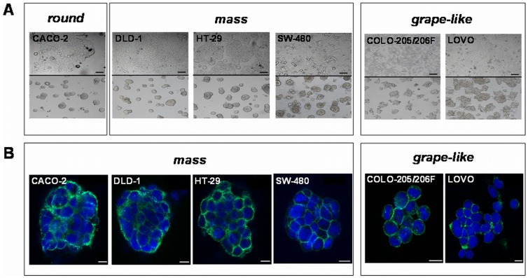 Fig. 1 Morphology of 2D and laminin-rich extracellular matrix (lrECM) 3D cultivated CRC cells. (A) Growth morphology of CRC cell lines cultivated under 2D (upper panel) and lrECM 3D on-top assay conditions (lower panel). Cells cultivated in 3D condition either show a round (CACO-2), mass (DLD-1, HT-29, SW-480) or a grape-like morphology (COLO 205, COLO-206F, LOVO) in phase contrast images. (B) Confocal laser scanning fluorescence microscopy images of CRC spheroids. (Luca AC, et al., 2013)
The division and proliferation of cancer cells are not terminated by cell contact with each other. Cells can accumulate to form a colony of cells when cultured in vitro, so cancer cell contact has no inhibitory effect on the proliferation of cancer cells.
Ask a Question
Average Rating: 5.0 | 1 Scientist has reviewed this product
Good
I'm so glad I chose the Creative Bioarray's products, it's the best!
12 Feb 2022
Ease of use
After sales services
Value for money
Write your own review
- You May Also Need

