ONLINE INQUIRY

Lobar Bronchial Epithelial Cells
Cat.No.: CSC-C4074X
Species: Human
Source: Bronchus
Cell Type: Epithelial Cell
- Specification
- Background
- Scientific Data
- Q & A
- Customer Review
The human lobar bronchial epithelial cells, that coat the inside of the lobar bronchi, form a vital part of the respiratory system. They form the first line of defense in the respiratory tract and are an essential part of healthy body functioning. These cells are usually arranged in a layer or layers of cuboidal or columnar epithelial cells, mostly consisting of ciliated cells, goblet (secretory) cells, and basal cells. Using coordinated cilia, ciliated cells snare inhaled debris and pathogens into mucus and propel them to the throat for expulsion. Goblet cells produce mucus, which engulfs the epithelial surface, thereby preventing infection. Basal cells in the basal membrane can divide and differentiate into other epithelial cells. If a lung is damaged, basal cells quickly multiply and differentiate to regenerate destroyed tissues, maintaining the integrity and functionality of the airway.
Lobar bronchial epithelial cells have distinct structural and morphological features in contrast to other bronchial epithelial cells. For example, epithelial cells closer to the lung hilum have larger cilia and a stronger mucus-secreting capacity to cope with increased exposure to bacteria and dust. Lobar bronchial epithelial cells respond distinctively to diverse environmental conditions, ensuring airway defense and normal functioning. As a result, these cells have found wide applications in science, especially in respiratory diseases. Through in vitro models, these cells can simulate the cellular mechanism of chronic obstructive pulmonary disease (COPD) and asthma, providing crucial insights into how disease operates.
 Fig. 1. Human lobar bronchus epithelium (Hollenhorst, M. I., Husnik, T., et al., 2023).
Pine WSPM Activates TRPV3 and TRPA1 in Lobar HBECs and Induces a Robust ERS Response
WSPM is a complex mixture of solids and condensed chemicals with variable pneumotoxic potential. Previous studies have demonstrated that WSPM from pine, mesquite, and other fuels differentially activate the transient receptor potential ankyrin-1 (TRPA1) and transient receptor potential vanilloid-3 (TRPV3) ion channels. However, the detailed mechanisms of how TRPA1 and TRPV3 ultimately influenced the cytotoxicity of WSPM have not been determined, leaving critical knowledge gaps surrounding the physiologic and pathophysiological functions of these channels in airway epithelial cells.
Nguyen's team investigated the roles of TRPA1 and TRPV3 in regulating endoplasmic reticulum stress (ERS) and cytotoxicity in human bronchial epithelial cells (HBECs) treated with WSPM and chemical agonists of each channel. Pine WSPM caused dose-dependent cytotoxicity with an LD50 of ∼18.8 μg/cm² (Fig. 1A). A short-term calcium flux assay indicated significant changes in cytosolic calcium following treatment with pine WSPM (78 μg/cm²) in lobar cells, which were partially inhibited by the TRPA1 antagonist A967079 (20 μM) and the TRPV3 antagonist (10 μM) (Fig. 1B). Pine WSPM activated both TRPA1 and TRPV3 in lobar HBECs, while neither antagonist alone triggered calcium flux. To evaluate the expression of ERS-associated biomarkers in lobar HBECs after pine WSPM treatment (20 μg/cm²), total mRNA was isolated at various times post-treatment. Significant time-dependent upregulation of PERK/ eIF2αK3 pathway biomarkers ATF3 and DDIT3 was observed, with transient HSPA1A elevation and sustained XBP1 mRNA splicing indicating IRE1α/β activation (Fig. 3). XBP1 splicing peaked within 2 hours, HSPA1A induction at 4 hours, and PERK-dependent ATF3/DDIT3 mRNA beyond 4 hours. Various WSPM materials also induced DDIT3 and ATF3, indicating ERS as a common response in lobar HBECs, with stronger TRPA1/TRPV3 agonists eliciting greater effects.
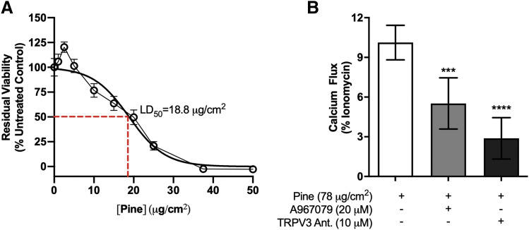 Fig. 1. WSPM cytotoxicity and the contributions of TRPV3 and TRPA1 in regulating pine WSPM–induced calcium flux (Nguyen ND, Memon TA, et al., 2020).
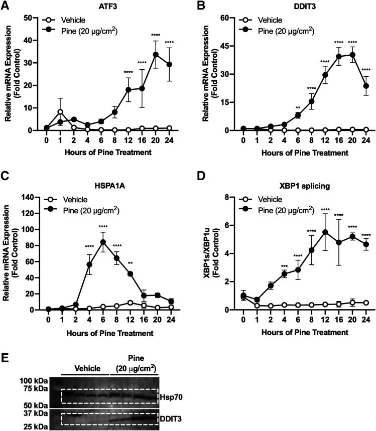 Fig. 2. Temporal profiling of ERS biomarkers in lobar HBECs in response to pine WSPM treatment (Nguyen ND, Memon TA, et al., 2020).
Pine WSPM Induced MUC5AC Gene Expression and Secretion in HLBECs
Previous studies have shown that combustion-derived particulate matter (PM) like wood smoke PM (WSPM) can cause excessive mucus due to transient receptor potential (TRP) channels activation, especially transient receptor potential ankyrin-1 (TRPA1). While TRPA1 partly mediates allergen-induced Muc5ac gene upregulation, other pathways like EGFR signaling also promote non-allergic MUC5AC overproduction, which is relevant in the case of WSPM. To further decipher mechanisms by which WSPM triggers nonallergic airway mucin gene expression and hypersecretion by HBECs. Pine WSPM and primary human lobar bronchial epithelial cells (HLBEC) were used as the main models.
HLBECs were treated with seven different size fractions of pine WSPM. Among the seven fractions tested, the PM2.5 and smaller fractions (F5, F6, and F7) significantly increased MUC5AC expression but did not significantly change MUC5B mRNA expression (Fig. 3A and B). The increase in MUC5AC mRNA expression by the pine WSPM F7 fraction was dose-dependent, with significant induction observed at concentrations of 20 μg/cm² or higher (Fig. 3C). Treatment with 20 μg/cm² of pine WSPM F7 resulted in about 40%±10% cell death within 24 hours, suggesting cell damage might contribute to MUC5AC gene expression. Longer treatments (48 hours) showed similar levels of mucin gene induction and increased cytotoxicity. Meanwhile, Muc5ac expression was also upregulated in the large airways of mice exposed to pine WSPM.
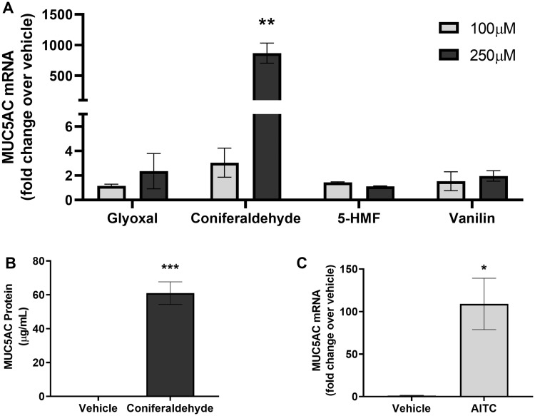 Fig. 3. Pine wood smoke particulate matter (WSPM) induced MUC5AC mRNA expression in human lobar bronchial epithelial cells and mice (Memon TA, Nguyen ND, et al., 2020).
It is recommended to use SuperCult® Bronchial Epithelial Cell Growth Basal Medium (cat# CM-1352L) and SuperCult® Bronchial Epithelial Cell Growth Meidum Supplement Kit (cat# CM-1353L) for the culturing of Lobar Bronchial Epithelial Cells.
Creative Bioarray’s Lobar Bronchial Epithelial Cells are isolated from the epithelial cells that line the airway of the bifurcation of the lungs and small sections of the bronchia just off of the bifurcation.
Primary Human Lobar Bronchial Epithelial Cells provide an ideal model for the studies of toxicity, cystic fibrosis, asthma, pathogenesis, pharmacology or airway wound healing.
Ask a Question
Average Rating: 5.0 | 1 Scientist has reviewed this product
Reliable
Creative Bioarray's Lobar Bronchial Epithelial Cells made a significant contribution to the success of our experiment.
18 Apr 2023
Ease of use
After sales services
Value for money
Write your own review


