ONLINE INQUIRY

Human Schwann Cells (HSC)
Cat.No.: CSC-7715W
Species: Human
Cell Type: Schwann Cell; Glial Cell
- Specification
- Background
- Scientific Data
- Publications
- Q & A
- Customer Review
During embryonic development neural crest cells migrate from the sides of the notochord towards the ectoderm before differentiating into multiple cell types including neurons and Schwann cells. Schwann cells develop into both myelinating and non-myelinating forms. While myelinating Schwann cells wrap axons in myelin sheaths to speed up nerve impulse transmission non-myelinating Schwann cells fulfill supportive roles without forming myelin. The origins of hSCs include adult neural tissues as well as developing neural tissues which contain adult spinal cords and sciatic nerves together with embryonic ganglia iPSCs and hESCs. hSCs exhibit a flat or polygonal shape with oval variations and possess noticeable nuclei along with a rich cytoplasm content.
These cells perform myelination functions while also assisting neural repair through regeneration and myelin restoration as well as managing immune responses by producing neurotrophic factors such as BDNF and GDNF. Human Schwann cells (hSCs) show tremendous potential for various applications including disease modeling of diabetic neuropathy and spinal cord injury research along with high-throughput drug testing for nerve regeneration and cell as well as gene therapy approaches for treating peripheral nerve and spinal cord injuries and genetic disorders through their use as delivery systems.
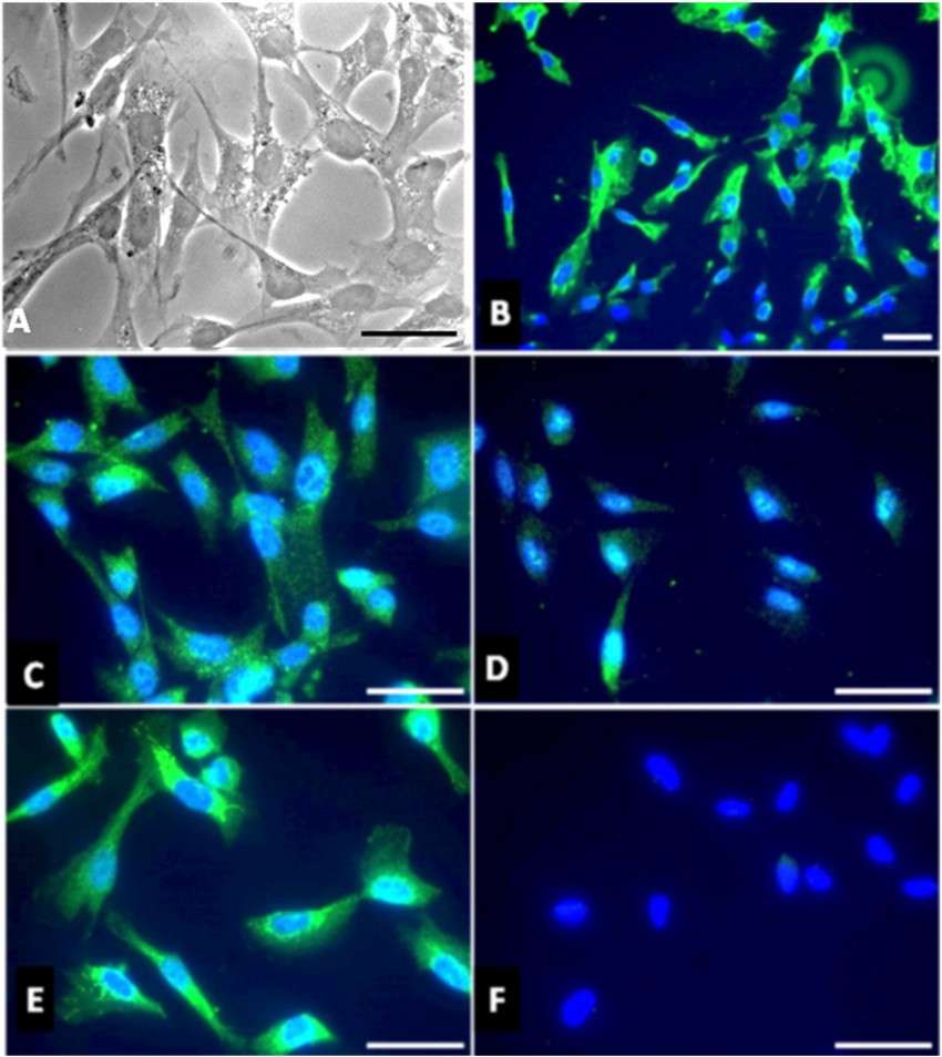 Fig. 1.Characterization of human Schwann cells in primary culture. Morphology of HSC in light microscopy (A). Immunocytofluorescence analyses of TLR4 (B), S100β (C), P0 myelin protein (D), and P75/NGF receptor (E). No staining is observed when the secondary antibody coupled to FITC is used alone (F). Cell nuclei are labelled with DAPI (blue), scale bar: 5 μm. (Faucard R, Madeira A, et al., 2016).
Low-Dose HT Increased the Viability and Proliferation of hSC
Phenolic compounds found in olive oil such as hydroxytyrosol (HT) deliver neuroprotective properties through antioxidative protection which recent phytomedicine studies emphasize to fight neurological disorders. Kamil et al. seek to understand how HT influences human Schwann cell (hSC) proliferation in vitro by evaluating modifications in growth speed and cellular growth protein markers.
To determine HT's effective dose, hSCs were treated with various concentrations for 24 hours. As shown in Figure 1A, cell morphology remained unchanged except at 50 and 100 µg/mL, where cells appeared round and shrunken. MTT assay results in Figure 1B indicated that 10 and 20 ng/mL HT significantly increased hSC proliferation with maintained viability (p < 0.05). One-way ANOVA confirmed the significant treatment effect (F (13,41) = 23.01; p < 0.0001). Higher concentrations, 50 and 100 µg/mL, reduced cell viability (p < 0.05). Thus, 20 ng/mL HT was selected for further experiments and referred to as "HT" henceforth. Low doses promote proliferation, while high doses inhibit it, similar to effects on human osteoblastic and endothelial cells.
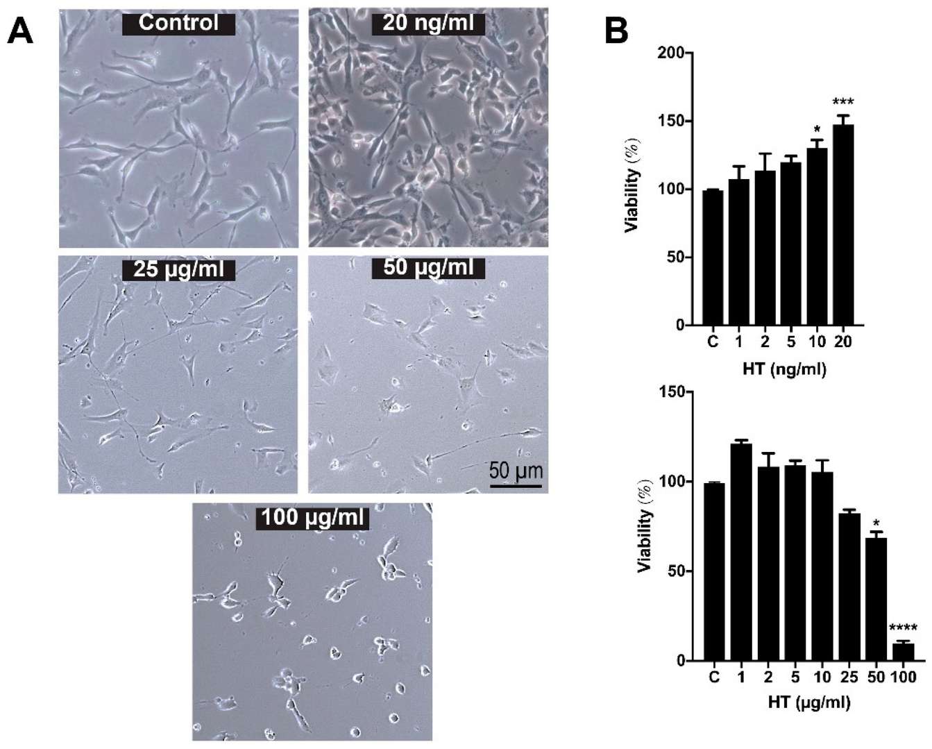 Fig. 1. Morphology and cell viability of hSC after 24-h treatment with hydroxytyrosol (HT) (Kamil K, Yazid MD, et al., 2020).
Functional Characterization of Muscarinic Receptors in Human Schwann Cells
The functional role of muscarinic receptors in myelinating glial cells has been established in rodent models, with M2 receptor subtype dominating in rat Schwann cells (SCs) and regulating proliferation/differentiation balance. However, human SCs (hSCs) remain understudied despite their clinical relevance to peripheral neuropathies. Piovesana et al. aimed to verify whether hSCs exhibit similar cholinoceptive properties to rodent SCs.
To assess muscarinic receptor modulation of hSC development, they analyzed cell growth via MTS assay in 5 hSCs using 100 µM APE or muscarine over 7 days (Fig. 2A). APE reduced cell growth by 72 h, remaining lower than controls at day 7. Conversely, muscarine initially decreased cell numbers at 3 days but promoted proliferation by day 5 (Fig. 2A). Supplementary analyses confirmed reduced growth in APE-treated versus control cells at all timepoint. To address the observed delayed APE effect (e.g., slight growth increase at day 7), they tested varying APE concentrations with media replacement at day 3. Here, 100 µM APE persistently suppressed growth at both 3 and 7 days (Fig. 2A). Only 50-100 µM APE significantly inhibited growth, with 50 µM effective only at day 7 (Fig. 2B). Similarly, muscarine enhanced proliferation solely at 100 µM (Fig. 2A).
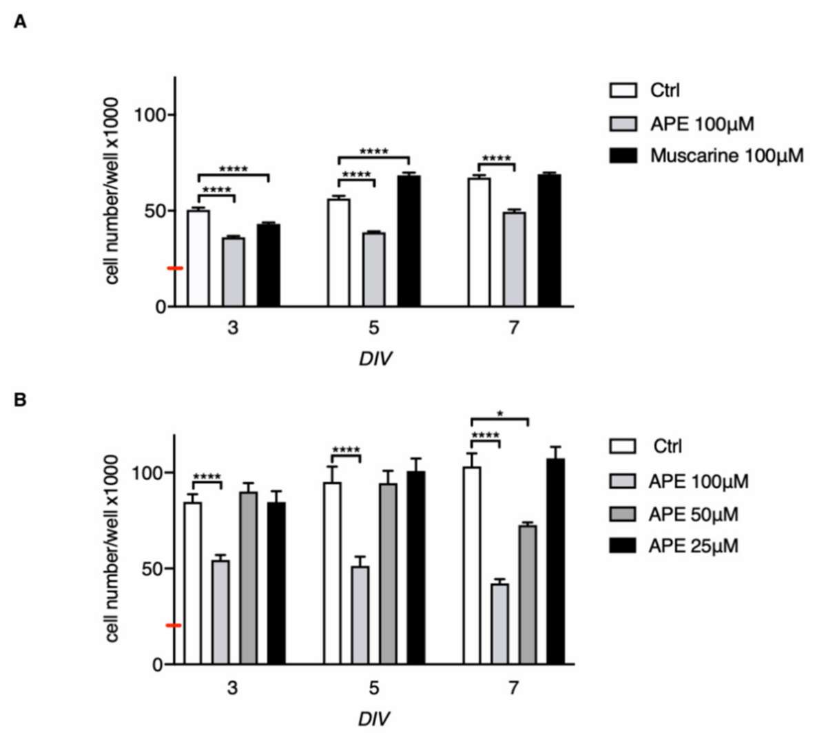 Fig. 2. MTS assay in hSCs maintained up to 7 days of treatment in vitro (DIV) in presence or absence of muscarinic agonists (Piovesana R, Faroni A, et al., 2020).
Yes, poly-L-lysine-coated culture vessel should be used to enhance human schwann cells attachement and adhesion. 2 μg/cm2 , T-75 flask is recommended.
Ask a Question
Write your own review
Description: Human Retinal Astrocytes (HRA) from Creative Bioarray are isolated from human retina. HRA are cryopreserved at passage one and delivered frozen. Each vial contains >5 x 10^5 cells in 1 ml volume. HRA are characterized by immunofluorescent method with antibody to GFAP. HRA are negative for HIV-1, HBV, HCV, mycoplasma, bacteria, yeast and fungi. HRA are guaranteed to further expand for 15 population doublings at the conditions provided by Creative Bioarray.
Description: Human Perineurial Cells (HPC) from Creative Bioarray Research Laboratories are isolated from human spinal nerves. HPNC are cryopreserved at passage one and delivered frozen. Each vial contains >5 x 10^5 cells in 1 ml volume. HPNC are characterized by immunofluorescent method with antibodies to Vimentin, S100, GFAP and CD90. HPNC are negative for HIV-1, HBV, HCV, mycoplasma, bacteria, yeast and fungi. HPNC are guaranteed to further expand for 15 population doublings in the conditions provided by Creative Bioarray.
Description: Human Oligodendrocyte Precursor Cells (HOPC) from Creative Bioarray are isolated from human brain tissue. HOPC are cryopreserved after purification and delivered frozen. Each vial contains >1 x 10^6 cells in 1 ml volume. HOPC are characterized by immunofluorescent method with antibodies to A2B5 and nestin. HOPC are negative for HIV-1, HBV, HCV, mycoplasma, bacteria, yeast and fungi. HOPC are guaranteed to further culture at the conditions provided by Creative Bioarray.
Description: Human Oligodendrocyte Precursor Cell-oligospheres (HOPC-os) from Creative Bioarray are isolated from human oligodendrocyte precursor cell cultures. HOPC-os are cryopreserved and delivered frozen. Each vial contains >1 x 10^6 cells in 1 ml volume. HOPC-os are characterized by immunofluorescent method with antibodies to A2B5 and nestin. HOPC-os are negative for HIV-1, HBV, HCV, mycoplasma, bacteria, yeast and fungi. HOPC-os are guaranteed to further culture at the condition provided by Creative Bioarray.
Description: Human Neurons-hippocampal (HN-h) from Creative Bioarray are isolated from hippocampal tissue of the brain. HHN are cryopreserved at primary cultures and delivered frozen. Each vial contains >1 x 10^6 cells in 1 ml volume. HHN are characterized by immunofluorescent method with antibodies to neurafilament, MAP2, and beta-tubulin III. HHN are negative for HIV-1, HBV, HCV, mycoplasma, bacteria, yeast and fungi. HHN are guaranteed to further culture in the conditions provided by Creative Bioarray.
Description: Human Neurons-brain stem (HMBS) from Creative Bioarray are isolated from human brain stem tissue. HN-bs are cryopreserved at primary culture and delivered frozen. Each vial contains >1 x 10^6 cells in 1 ml volume. HN-bs are characterized by immunofluorescence with antibodies specific to neurofilament, MAP2, and beta-tubulin III. HN-bs are negative for HIV-1, HBV, HCV, mycoplasma, bacteria, yeast and fungi. HN-bs are guaranteed to further culture in the conditions provided by Creative Bioarray.
