ONLINE INQUIRY

Human Osteoblast Cells
Cat.No.: CSC-7860W
Species: Human
Source: Bone
Cell Type: Osteoblast
- Specification
- Background
- Scientific Data
- Q & A
- Customer Review
In humans, osteoblasts mainly come from the bone, the periosteum, the bone marrow and extra-skeletal tissues. Creative Bioarray's human osteoblast cell lines come from trabecular bone tissue of the knee or hip joint. These cells originate from mesenchymal cells or bone marrow stromal cells that possess multipotent differentiation abilities. They descend from fibroblasts and chondrocytes, which together comprise an elaborate cell family tree. Osteoblasts usually lie on the surface of the bone, in different shaped (cuboidal, short columnar, spindle) close together to form a protective coating. These cells under an optical microscope exhibit massive, round nuclei, basophilic cytoplasm and thick coarse endoplasmic reticulum and Golgi apparatus, suggesting metabolic activity.
Osteoblasts function primarily to make bone matrix, manage bone matrix mineralisation and affect osteoclast bone resorption. They stimulate bone growth and repair by continually producing and releasing bone matrix proteins. Further, osteoblasts coordinate bone remodeling and regrowth by releasing cytokines and growth factors, keeping bones healthy and robust. For research, osteoblasts are used to investigate bone metabolism, treatment of bone disease and the engineering of bone tissue. Such research contributes to an increased awareness of bone physiology, to the creation of novel therapies, and to the development of bone tissue engineering.
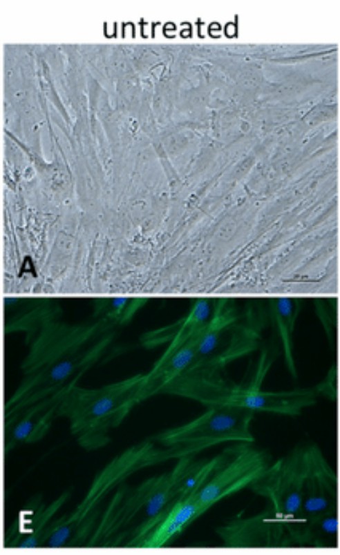 Fig. 1. Cell morphology of human osteoblasts. (A) Phase contrast microscopy (scale bar: 20 µm); (E) Fluorescence staining of actin filaments and cell nuclei (scale bar: 50 µm) (Jonitz-Heincke A, 2019).
EFMF Induced Proliferation and Mineralization of Primary Osteoblasts In Vitro
In vertebrates, bone is formed by intramembranous and endochondral ossification, the latter being responsible for healing fractures. Endochondral ossification involves mesenchymal cells becoming osteoblasts and depositing bone matrix over cartilage. Previous research demonstrated that magnetic field and an additional electric field (EFMF) treatment promoted cell differentiation and protein production, suggesting potential effects on bone healing.
To evaluate the molecular effects of combined exposure to EF and MF, Dittmann's team exposed human primary osteoblasts (hOBs) under standardized conditions to a combination of an EF (20 Hz, 700 mV) and a MF (20 Hz, 5 mT). Figure 1 shows primary osteoblast population doublings over 21 days, where cell proliferation was notably enhanced by about 25% up to day 14 under EFMF exposure. To prevent contact inhibition, cells were initially seeded at reduced numbers. Alizarin Ca2+ staining indicated an increase in Ca2+ deposition (1.11), further amplified with EFMF to 1.44 (Fig. 3), alongside elevated expression of Ca2+-related genes Catherin11 (1.40) (CDH11) and cartilage oligomeric matrix protein (COMP) (1.50). The observed changes in phenotype and the increased Ca2+ deposition in EFMF-exposed osteoblasts indicated an induced differentiation process of the applied hOBs. To characterize the associated functional alterations, we summarized the protein expression (Fig. 2) of essential genes involved in osteoblast differentiation and function that responded to EFMF.
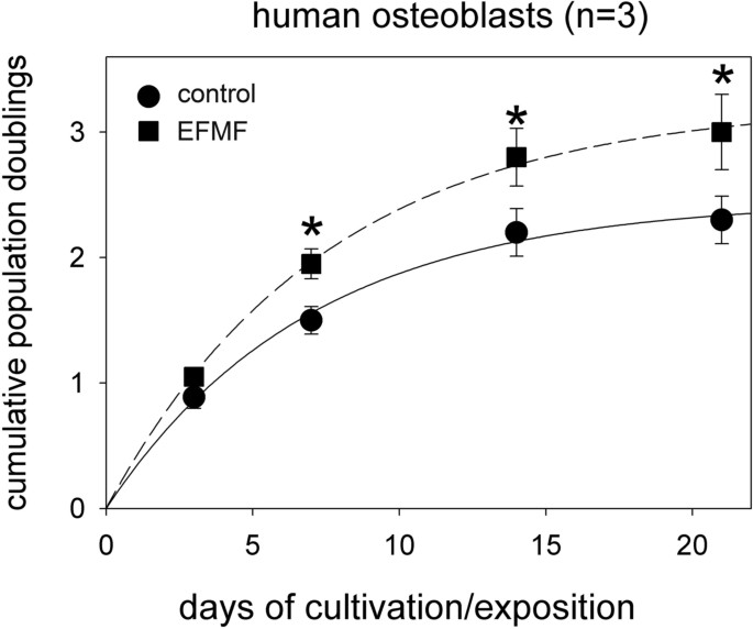 Fig. 1. Effect of EFMF exposure on cell proliferation of hOBs (Dittmann KH, Mayer C, et al., 2022)
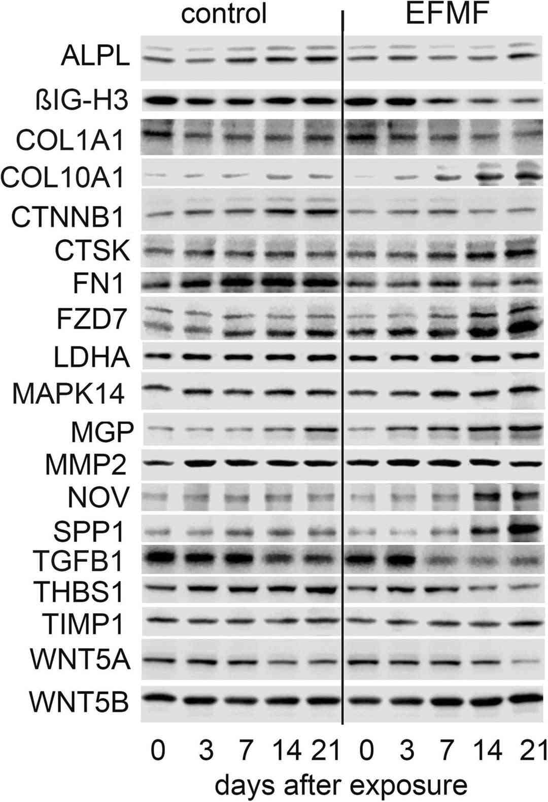 Fig. 2. Time-dependent protein expression of selected proteins of hOBs in response to EFMF treatment (Dittmann KH, Mayer C, et al., 2022).
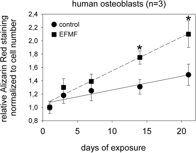 Fig. 3. Alizarin red staining of hOBs after exposure to EFMF (Dittmann KH, Mayer C, et al., 2022).
Evaluation of Ethanol-Induced Chitosan Aerogels with Human Osteoblast Cells
Chitosan, a natural polysaccharide, offers potential as a biomaterial due to its biocompatible and biodegradable properties. Traditionally, chitosan aerogels are synthesized using chemical cross-linkers, but these can be toxic. Recent trends focus on using natural materials for green chemistry solutions.
Pantić et al. explored ethanol-based synthesis of chitosan aerogels without external cross-linkers. Then, they tested the biocompatibility and cytotoxicity of this chitosan aerogels with human osteoblasts through an extraction/elution assay. Human osteoblasts were treated with serial dilutions for 24 h, and using the MTT assay to assess the cell viability. It was found that extracts from pure chitosan aerogels did not affect cell viability (Fig. 4a). In contrast, extracts from ethanol/NaOH-prepared aerogels decreased cell viability for both undiluted and 1:2 dilutions. As shown in Fig. 4b, cells adhered and survived on the ethanol-prepared chitosan aerogels, indicated by green fluorescence. Conversely, red staining on aerogels prepared with ethanol/NaOH pointed to cell death. Additionally, immunocytochemical analysis was used to detect osteocalcin, an osteoblast marker, in human osteoblasts cultured on ethanol-prepared chitosan aerogels for seven days. Cell viability (Fig. 5a) and osteocalcin expression were confirmed (Fig. 5).
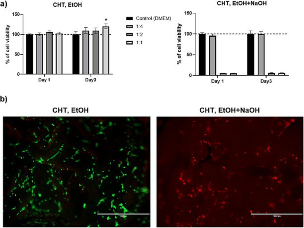 Fig. 4. Viability of HOB cells when treated with chitosan aerogel elution (a) and direct contact test using a live/dead assay (b) (Pantića M, Maver U, et al., 2023).
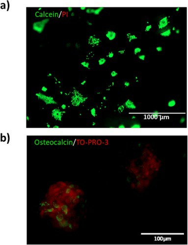 Fig. 5. Cell viability and immunofluorescence for osteocalcin in HOB cells after seven days of culture (Pantića M, Maver U, et al., 2023).
Creative Bioarray’s Human Osteoblasts are isolated by explant, and the tissue is from the spongy section of the bone head of the ribs, femur, or tibia.
No, our osteoblast cells are not contact inhibited and tend to grow on top of each other after becoming confluent.
The average doubling time of our osteoblasts is 35-40 hours in earlier passages. If in older (later passage) cultures, it can be much longer, and there might be donor-to-donor variations as well.
We have found some osteoblast cell strains to be more sensitive to β-Glycerophosphate than others. Therefore, we recommend trying two cited concentration to determine the optimal concentration for each specific lot.
Ask a Question
Average Rating: 5.0 | 1 Scientist has reviewed this product
So fast and helpful
The Creative Bioarray Team has been extremely helpful and patient in assisting me in placing orders for cells and recommending appropriate media and protocols. Any orders I have placed have arrived faster than expected and have grown without issue.
12 May 2022
Ease of use
After sales services
Value for money
Write your own review
Description: Human Osteoblasts-femural (HO-f) from Creative Bioarray are isolated from human femur. HO-f are cryopreserved at primary cultures and delivered frozen. Each vial contains >5 x 10^5 cells in 1 ml volume. HO-f are characterized by the cytochemically detection of AP and mineral deposition. HO-f are negative for HIV-1, HBV, HCV, mycoplasma, bacteria, yeast and fungi. HO-f are guaranteed for 15 population doublings at the conditions provided by Creative Bioarray.
Description: Human Calvarial Osteoblasts (HCO) from Creative Bioarray are isolated from human calvariae. HCO are cryopreserved at primary cultures and delivered frozen. Each vial contains >5 x 10^5 cells in 1 ml volume. HCO are characterized by the cytochemically detection of AP and mineral deposition. HCO are negative for HIV-1, HBV, HCV, mycoplasma, bacteria, yeast and fungi. HCO are guaranteed for 15 population doublings at the conditions provided by Creative Bioarray.
