- HCC-78
- HDLM-2
- DOHH-2
- L-540
- MX-1
- NALM-6
- NB-4
- CAL-51
- SNB-19
- KYSE-520
- MKN-45
- BA/F3
- MS-5
- HCEC-B4G12
- NK-92
- PA-TU-8988S
- MONO-MAC-1
- PA-TU-8902
- Human Microglia
- Human Hepatic Stellate Cells
- Human Skeletal Muscle Cells (DMD)
- Human Schwann Cells
- Human Oral Keratinocytes (HOK)
- Human Cardiomyocytes
- Human Small Intestinal Epithelial Cells
- Human Colonic Epithelial Cells
- Human Intestinal Fibroblasts
- Primary Human Large Intestine Microvascular Endothelial Cells
- Human Small Intestinal Microvascular Endothelial Cells
- Human Retinal Pigment Epithelial Cells
- Human Hepatocytes
- Cynomolgus Monkey Lung Microvascular Endothelial Cells
- Cynomolgus Monkey Vein Endothelial Cells
- C57BL/6 Mouse Primary Mammary Epithelial Cells
- C57BL/6 Mouse Vein Endothelial Cells
- Rat Primary Kidney Epithelial Cells
- Rat Gingival Epithelial Cells
- Rabbit Lung Endothelial Cells
Our Promise to You
Guaranteed product quality, expert customer support
ONLINE INQUIRY
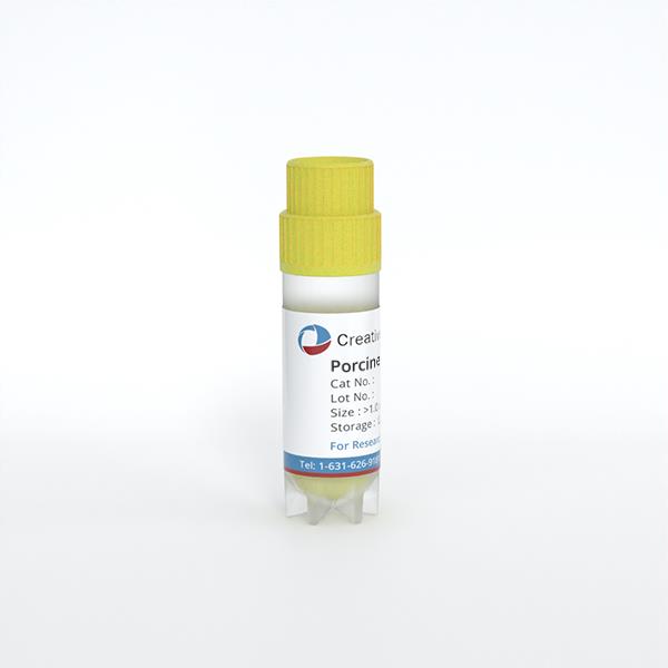
Porcine Spleen Epithelial Cells
Cat.No.: CSC-C9254J
Species: Porcine
Source: Spleen
Cell Type: Epithelial
- Specification
- Q & A
- Customer Review
Once you find a black spot in the process of cell culture, firstly, observe the culture medium with the naked eye to see if it is cloudy, and then, under the microscope, observe the state of the cultured cell growth and whether the black spot is swimming. If the cells are contaminated, the bacteria will multiply and the medium will rapidly turn yellow and cloudy.
Ask a Question
Average Rating: 5.0 | 1 Scientist has reviewed this product
Excellent results
I have used these cells several times and have had excellent experimental results.
23 Apr 2023
Ease of use
After sales services
Value for money
Write your own review