Description
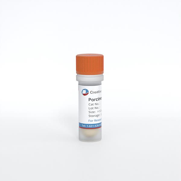
Porcine Lung Microvascular Endothelial Cells from Creative Bioarray are isolated from lung tissue of porcine. Porcine Lung Microvascular Endothelial Cells are grown in T25 tissue culture flasks pre-coated with gelatin-based coating solution for 2 min and incubated in Creative Bioarray’ Culture Complete Growth Medium generally for 3-7 days. Cultures are then expanded. Prior to shipping, cells are detached from flasks and immediately cryo-preserved in vials. Each vial contains at least 0.5x10^6 cells per ml and are delivered frozen. The method we use to isolate endothelial cells was developed based on a combination of established and our proprietary methods. These cells are pre-coated with PECAM-1 antibody, following the application of magnetic pre-coated with secondary antibody.
Cell Type
Endothelial Cell; Microvascular Cell
Quality Control
Porcine Lung Microvascular Endothelial Cells are tested for uptake of Dil-Ac-LDL (Catalog No. L-35353, Invitrogen), a functional marker for endothelial cells. Porcine Lung Microvascular Endothelial Cells are negative for bacteria, yeast, fungi and mycoplasma. Cells can be expanded for 3-5 passages at a split ratio of 1:2 under the cell culture conditions specified by Creative Bioarray. Repeated freezing and thawing of cells is not recommended.
Storage and Shipping
Creative Bioarray ships frozen cells on dry ice. On receipt, immediately transfer frozen cells to liquid nitrogen (-180 °C) until ready for experimental use. Live cell shipment is also available on request.
Never can primary cells be kept at -20 °C.
Citation Guidance
If you use this products in your scientific publication, it should be cited in the publication as: Creative Bioarray cat no.
If your paper has been published, please click here to submit the PubMed ID of your paper to get a coupon.
How should cell contamination be avoided?
The types of cell contamination can be classified as bacteria, yeast, mold, virus and mycoplasma. The main causes of contamination are improper aseptic technique, poor operating room environment, contaminated serum and contaminated cells. Strict aseptic technique, clean environment, good quality cell source and medium preparation are the best ways to reduce contamination.


