ONLINE INQUIRY
Human Small Intestinal Epithelial Cells
Cat.No.: CSC-C9229J
Species: Human
Source: Small Intestine; Intestine
Cell Type: Epithelial Cell
- Specification
- Background
- Scientific Data
- Publications
- Q & A
- Customer Review
Human small intestinal epithelial cells (IECs) are highly specialized cells that line the lumen of the small intestine, playing key roles in nutrient absorption, barrier function, and immune regulation. These cells form a monolayer epithelium organized into crypt-villus structures, which maximize the surface area for efficient nutrient uptake. Functionally, IECs are responsible for the digestion and absorption of carbohydrates, proteins, and fats by expressing specific transporters and enzymes, such as disaccharidases and peptidases. Additionally, they maintain the integrity of the intestinal barrier via tight junctions, preventing the translocation of pathogens and toxins. IECs also contribute to mucosal immunity by secreting antimicrobial peptides (e.g., defensins from Paneth cells), mucus (from goblet cells), and cytokines, while interacting with immune cells to modulate local and systemic immune responses. Some of the most severe pathological conditions, such as inflammatory bowel disease (IBD), are known to be associated with alterations of the normal growth and function of the intestinal epithelium. It is therefore of interest to investigate the physiology and pathophysiology of these cells and, not surprisingly, a large number of scientists in research fields such as intestinal physiology, intestinal immunology and cancer genesis are interested in the study of these cells.
Advantages of using human small intestinal epithelial cells include their relevance to human biology, the ability to mimic in vivo conditions, and the versatility in experimental setups. However, challenges remain, such as maintaining long-term viability and functional stability in vitro. Advances in 3D culture systems and organ-on-a-chip technologies are addressing these limitations, further enhancing their utility in biomedical research.
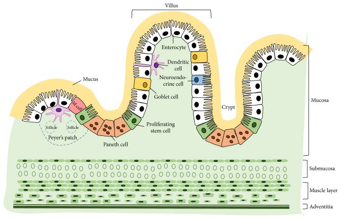 Fig. 1. Cross-sectional structure of small intestine and the major cell constituents of epithelium (Kong, Shanshan, et al. 2018).
Anti-Inflammatory Effects in Intestinal Epithelial Cells
Given growing understanding of the importance of peripheral inflammation, gut-health and the gut-brain axis on cognitive health, the effects of S. officinalis extract were assessed in human small intestinal epithelial cells as a model of the intestinal barrier.
Figures 1 and 2 summarize the levels of cytokines and chemokines released into apical and basolateral cell media respectively by human small intestinal epithelial cells incubated with LPS and S. officinalis extract for 24 h. Compared to the control group, the LPS treatment caused a significant increase in the release of all eight of the protein markers tested in the apical media (Fig. 1) and all but CRP and VCAM-1 in the basolateral media (Fig. 2), which supports successful induction of pro-inflammatory response by the LPS treatment.
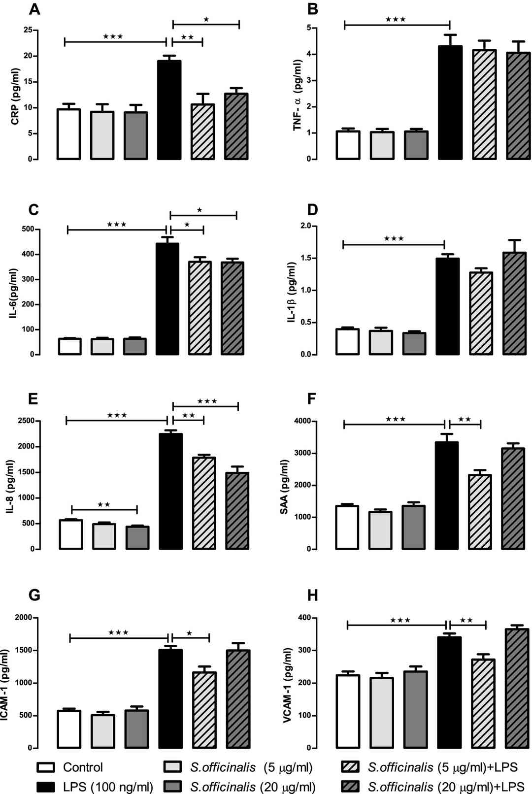 Fig. 1. S. officinalis extracts attenuation of cytokine and chemokine release by human small intestinal epithelial cells in apical media after 24-h treatment with LPS (Margetts, Gemma, et al. 2022).
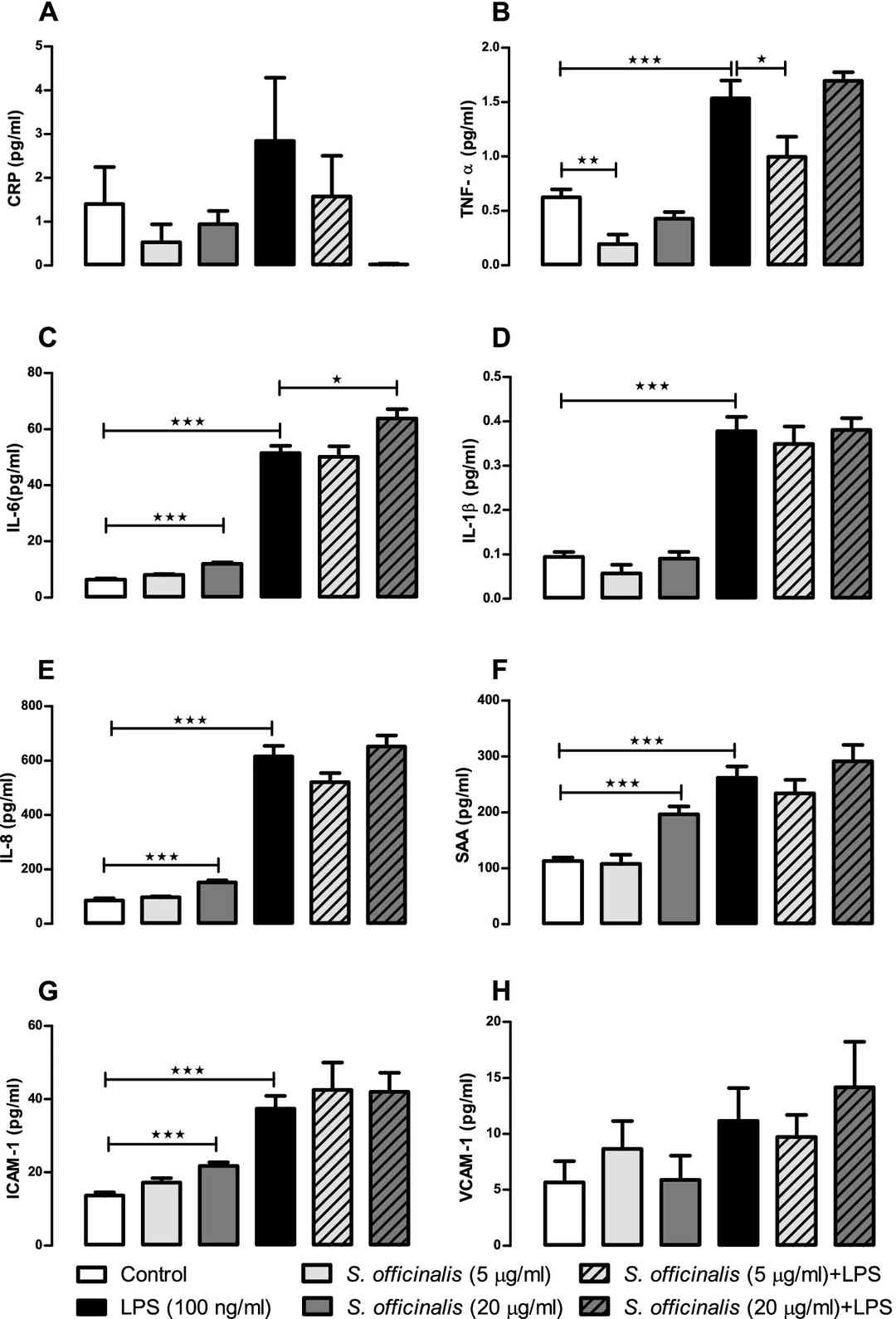 Fig. 2. S. officinalis extracts attenuation of cytokine and chemokine release by human small intestinal epithelial cells in basolateral media after 24-h treatment with LPS (Margetts, Gemma, et al. 2022).
Characterization of Conditions for The Primary Culture of Human Small Intestinal Epithelial Cells
Intestinal epithelial cells (IECs) were cultured on collagen membranes in a 12-well tissue culture cluster, with Lamina propria (LP) cells and allogeneic Epstein-Barr Virus (EBV)-transformed B lymphocytes (allo-B cells) underneath, in the well.
Primary human IECs in culture did not form confluent monolayers but cells did maintain contact with other cells in small sheets. Individual cells did maintain their cellular polarity but the cells were not all aligned on the membrane.
Initial experiments of IECs cultured above the LP fraction and allo-B cells (E/LP + B), showed that cells cultured for one day maintained their columnar/cuboid morphology and polarity (Fig. 3a, b). Morphology was further maintained when cells were cultured for 2 days (Fig. 3c, d) and for 7 days (Fig. 3f, g). All IECs expressed the human epithelial antigen as detected by Ber-EP4 antibody (Fig. 3f, g), and there was no evidence of contamination by fibroblasts.
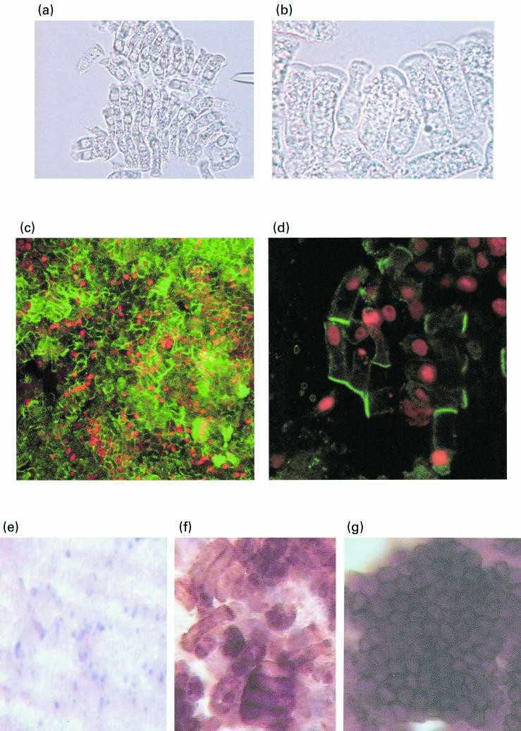 Fig. 3. Culture of epithelial cells above LP fraction and allo-B cells in the culture system (Aldhous, M. C., et al. 2001).
Our cells can be handled at biosafety level 1, but caution is always recommended when handling human tissue derived products. Hope this helps.
Ask a Question
Write your own review

Description: Smooth muscle contraction is the fundamental event in gastrointestinal motion. Inflammation of the human intestine causes thickening of the smooth muscle layers which results from the increases in the smooth muscle-specific actins. The increased smooth muscle actins may affect force production and further demonstrate the plasticity of smooth muscle in the inflamed intestine. Human intestinal smooth muscle cells respond to IL-1-beta and TNF-alpha stimulation by releasing IL-6, which might significantly contribute to the overall systemic inflammatory response. Knowledge of molecular mechanism that underlies the control of colorectal smooth muscle tone is essential to advance of understanding of pathophysiology of the abnormality. The availability of human rectal smooth muscle cells in culture will considerably enhance our ability to study the contractile, proliferative and connective tissue responses of the smooth muscle of the human colorectal disorders.HRSMC from Bioarray Research Laboratories are isolated from the human rectum. HRSMC are cryopreserved at primary or passage one culture and delivered frozen. Each vial contains >5 x 10^5 cells in 1 ml volume. HRSMC are characterized by immunofluorescent method with antibodies to Α-smooth muscle actin and desmin. HRSMC are negative for HIV-1, HBV, HCV, mycoplasma, bacteria, yeast and fungi. HRSMC are guaranteed to further expand for 15 population doublings in the condition provided by Bioarray Research Laboratories.
Description: Smooth muscle contraction is the fundamental event in gastrointestinal motion. Although many of the biochemical mechanisms underlying the excitation-contraction coupling are not yet defined, it is known that cytosolic Ca2+ is the essential component in the coupling phenomenon. Inflammation of the human intestine causes thickening of the smooth muscle layers which results from the increases in the smooth muscle-specific actins. The increased smooth muscle actins may affect force production and further demonstrate the plasticity of smooth muscle in the inflamed intestine. Studies also show that human intestinal smooth muscle cells respond to IL-1beta and TNF-alpha stimulation by releasing IL-6, which might significantly contribute to the overall systemic inflammatory response. The availability of human intestinal smooth muscle cells in culture will considerably enhance our ability to study the contractile, proliferative and connective tissue responses of the smooth muscle of the human gastrointestinal tract.HISMC from Bioarray Research Laboratories are isolated from human intestine. HISMC are cryopreserved at passage one culture and delivered frozen. Each vial contains >5 x 10^5 cells in 1 ml volume. HISMC are characterized by immunofluorescent method with antibodies to Α-smooth muscle actin and desmin. HISMC are negative for HIV-1, HBV, HCV, mycoplasma, bacteria, yeast and fungi. HISMC are guaranteed to further expand for 15 population doublings in the condition provided by Bioarray Research Laboratories.
Description: Endothelial cells lining the microvasculature are known to play a critical "gatekeeper" role in the inflammatory process through their ability to recruit circulating immune cells into tissues and foci of inflammation. Studies show that intestinal microvascular endothelial cells (IMEC) exhibit a strong immune response to LPS challenge and play a critical regulatory role in gut inflammation. Pharmacological inhibition of NOS in activated HIMEC resulted in a significant increase in leukocytes binding. Gene expression profile study reveals that intestinal endothelial cells express biotinidase, which is involved in biotin recycling. The in vitro culture of HIMEC enabled scientists to perform systematic analyses of the cytokine profiles with regard to mRNA expression and protein secretion, and to compare such data with cytokine profiles concomitantly displayed by other endothelial cells.HIMEC from Bioarray Research Laboratories are isolated from human intestinal tissue. HIMEC are cryopreserved at passage one and delivered frozen. Each vial contains >5 x 10^5 cells in 1 ml volume. HIMEC are characterized by immunofluorescent method with antibodies to vWF/Factor VIII and CD31 (P-CAM) and by uptake of DiI-Ac-LDL. HIMEC are negative for HIV-1, HBV, HCV, mycoplasma, bacteria, yeast and fungi. HIMEC are guaranteed to further culture in the conditions provided by Bioarray Research Laboratories.
Description: Fibroblasts are mesenchymal cells derived from the embryonic mesoderm. They have been extensively used for a wide range of cellular and molecular studies. This is mainly because they are one of easiest types of cells to grow in culture, and their durability makes them amenable to a wide variety of manipulations ranging from studies employing gene transfection to microinjection. There is good evidence that fibroblasts in different parts of the body are intrinsically different. Fibroblasts secrete a non-rigid extracellular matrix that is rich in type I and/or type III collagen. They are responsible for much of the synthesis of extracellular matrix in connective tissues and play major roles in wound healing. Many diseases are associated with fibroblasts, either because fibroblasts are implicated in their etiology or because of the fibrosis that accompanies damage to other cell types in tissues. For example, the development of bowl stenosis in Crohn's disease patients is caused by extreme fibroblast proliferation and extracellular matrix expansion.
HIF are isolated from human intestinal tissue. HIF are cryopreserved at passage one and delivered frozen. Each vial contains >5 x 10^5 cells in 1 ml volume. HIF are characterized by their spindle morphology and immunofluorescent method with antibody to fibronectin. HIF are negative for HIV-1, HBV, HCV, mycoplasma, bacteria, yeast and fungi.
Description: Smooth muscle is responsible for the contractility of hollow organs, such as blood vessels, the gastrointestinal tract, the bladder, and the uterus. Its structure differs greatly from that of skeletal muscle. The human stomach contains three layers of muscle in its walls, the outer longitudinal, the middle circular and the inner oblique and visceral smooth muscle cells makes up all three layers along the entire organ. Smooth muscle contraction is critical to peristalsis in the human stomach and the contraction may be mediated by activation of phospholipase through two distinct mechanisms (increased intracellular Ca2+ and G protein activation) and activating PKCepsilon-dependent mechanisms. In vitro study also shows that gastric smooth muscle cells express ET and eNOS and both calcium and sodium may be involved as current carriers in the generation of the plateau potential.HGSMC from Bioarray Research Laboratories are isolated from the human stomach. HGSMC are cryopreserved at secondary culture and delivered frozen. Each vial contains >5 x 10^5 cells in 1 ml volume. HGSMC are characterized by immunofluorescent method with antibodies to Α-smooth muscle actin and desmin. HGSMC are negative for HIV-1, HBV, HCV, mycoplasma, bacteria, yeast and fungi. HGSMC are guaranteed to further expand for 15 population doublings at the condition provided by Bioarray Research Laboratories.
Description: Primary Human Intestinal Microvascular Endothelial Cells were initiated by elutriation from dissociated normal human small intestine tissue.
These cells were originated using Complete Serum-Free Medium Kit With SuperFuel™, are available at <12 Cumulative Population Doublings (CPD) in vitro [Passage 3] and were cryopreserved in aliquots of ~1.5 X 10^6 cells. This vial will initiate a Passage 4 cell culture in a 75cm2 flask.
