ONLINE INQUIRY

Human Epidermal Melanocytes-light (HEM)
Cat.No.: CSC-7786W
Species: Human
Source: Epidermis; Skin
Cell Type: Melanocyte
- Specification
- Background
- Scientific Data
- Q & A
- Customer Review
Human Epidermal Melanocytes-light (HEM-l) are a type of lightly pigmented melanocytes derived from neonatal epidermal tissue, playing a distinct role in skin pigmentation and physiological function. These cells exhibit a multipolar or dendritic morphology, characterized by needle-like appearances, and predominantly adherent growth in the medium. Although morphologically similar to HEM-dark cell lines, they typically contain fewer melanin granules. Located in the basal layer of the epidermis, a hub of active skin cell renewal and pigment synthesis, they facilitate communication and transfer of signals and substances with surrounding keratinocytes via their dendritic structures.
Despite the basic functional similarities with HEM-dark cell lines, HEM-l cells have limited UV defense capabilities due to lower melanin content. Nonetheless, they are crucial in the formation of skin color. The unique biological responses of HEM-l cells to sunlight exposure and skin damage make them an ideal model for studying UV reactions and skin diseases such as psoriasis and vitiligo. Furthermore, HEM-l cells are critical in the development of cosmetic products, providing essential scientific validation in the efficacy assessments of skincare solutions, particularly in whitening and sunscreen products.
 Fig. 1. Human epidermal melanocytes from children's foreskin loaded with the Ca2+ indicator Fluo-3 (Lan Y, Zeng W, et al., 2021).
Effect of Bepridil on Melanocytes Cells
Research into calcium ion (Ca2+) signalling in melanoma has largely dealt with the Ca2+ entry mechanism, and has little concern for Ca2+ extrusion through the Na+/Ca2+ exchanger (NCX). Liu's team studies whether pharmacological regulation of the NCX can prevent the proliferation of melanoma, testing various inhibitors to look at their effect on Ca2+ and cell growth.
Liu's team indicated that Bepridil can induce Ca2+ related melanoma cell death by blocking the forward operation of NCX. Subsequently, they used melanocytes (HEM-L) and skin fibroblasts (HSF) as normal cell counterparts to melanoma and treated them with Bepridil, assessing their cell viability. They found that A2058, A375, and C8161 melanoma cells showed a significant reduction in viability (43-88%), whereas HEM-L and HSF cells were less affected (11-18%) (Fig. 1A). By Western blot, melanoma cells expressed more NCX1 isoform than HEM-L cells (Fig. 1B and C), extended to SK-MEL-2, SK-MEL-28, WM-115, and A875 (Fig. 2A-C and D). Bepridil killed A875, WM-115 and SK-MEL-28 cells in dose-dependent ways, yielding IC50 values of 1.60, 8.70 and 13.64 M, respectively (Fig. 2E).
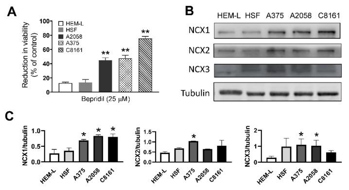 Fig. 1. The impact of bepridil on melanocytes cell (Liu Z, Cheng Q, et al., 2022).
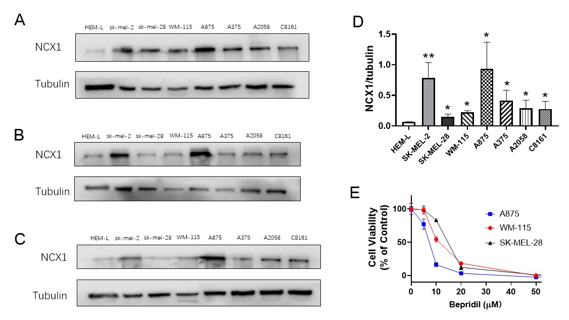 Fig. 2. Western blotting analysis of samples from eight different cell lines (Liu Z, Cheng Q, et al., 2022).
Fucoxanthin is Cytotoxic to Human Melanoma Cell Lines while Sparing Normal Human Epidermal Melanocytes
The most lethal skin cancer, melanoma, is seeking new treatments. The marine carotenoid fucoxanthin, too, is an anticancer agent – although it is unknown whether it inhibits human melanoma. In order to establish whether Fucoxanthin is effective against melanoma, Kuo et al. assessed fucoxanthin's cytotoxicity in human epidermal melanocytes-light (HEM-L) and normal human melanoma cells (A2058 and A375). These findings demonstrated that Fucoxanthin was cytotoxic in different concentrations in the human melanoma cell lines A2058 and A375, and slowed cell death (IC50 = 22-44 M for both cell lines) 24 to 48 hours following exposure. For clonogenicity tests, Fucoxanthin significantly lowered both melanoma cell lines' colony-forming capacities to approximately 33%, at 50 M concentration. In contrast, HEM-L were much less affected, retaining viability even at 50 μM for 24 hours, while dropping to 50% after 48 hours (Fig. 3). These results underscore Fucoxanthin is cytotoxic to human melanoma cell lines A2758 and A375 while showing limited cytotoxicity to normal human melanocytes.
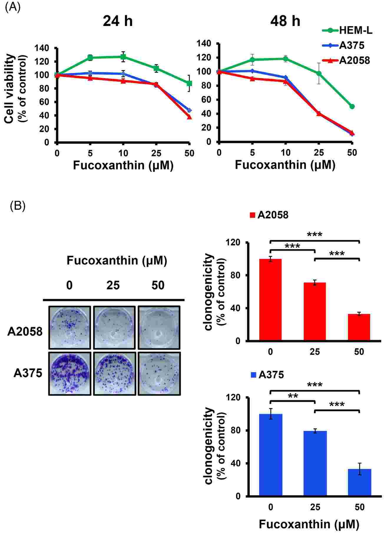 Fig. 3. Fucoxanthin is cytotoxic to human melanoma cells while sparing normal human melanocytes (Kuo MY, Dai WC, et al., 2024).
Ask a Question
Write your own review

