ONLINE INQUIRY

Human Podocyte
Cat.No.: CSC-C0320Z
Species: Human
Source: Kidney
Cell Type: Podocyte
- Specification
- Background
- Scientific Data
- Q & A
- Customer Review
The human podocyte cell line is a key in vitro model for studying human glomerulopathies. The cell bodies are usually round or oval and have a huge nucleus. Numerous elongated foot processes emerge from the cell wall, twisting and joining to create a branch-like structure. These foot processes are covered by complexes of proteins that adhere to the GBM via adhesion molecules and proteoglycans. In the foot processes, there are many 30-40 nanometre slits and between the slits 4-6 nanometer-thick slit diaphragms define the last obstacle to plasma proteins entering the vascular system.
Podocytes can also secrete extracellular matrix materials, such as collagen and laminin, and aid in extracellular matrix degradation. In accurately balancing synthesis and degradation of the extracellular matrix, podocytes preserve the stability and proper functioning of the glomerular basement membrane. In renal physiology, podocytes receive and broadcast signals, adapt to hemodynamics, and regulate their activity via a cascade of intracellular signaling networks. Podocytes also communicate with other glomerular cells, including endothelial and mesangial cells, to help collectively control the homeostasis of the glomerulus. Since podocyte structural and functional abnormalities can cause proteinuria and hematuria in glomerular disease, detailed studies of the signal transduction and gene expression regulation of Human Podocyte cells are essential to discovering novel treatments for kidney disease.
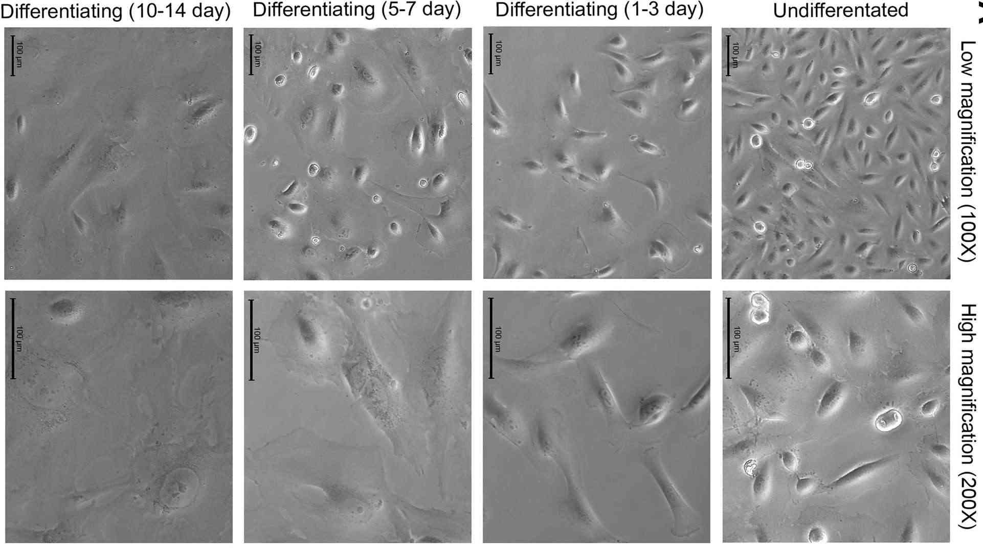 Fig. 1. Identification of differentiated human immortalized podocytes (Martinez-Arroyo O, Ortega A, et al., 2020).
Tacrolimus Prevents TWEAK-Induced PLA2R Expression in Cultured Human Podocytes
Primary membranous nephropathy (MN) is the leading cause of nephrotic syndrome among nondiabetic Caucasian adults and is usually caused by antibodies against the podocyte antigen membrane M-type phospholipase A2 receptor (PLA2R). While calcineurin inhibitors like tacrolimus are standard treatments, they often lead to disease recurrence upon withdrawal. In this regard, whether calcineurin inhibitors modulate podocyte PLA2R expression has not been explored so far. Cuarental's team investigated the factors influencing PLA2R expression in podocytes and the impact of tacrolimus especially focusing on the TNF receptor superfamily member TNFRSF12a (Fn14) and its ligand TWEAK.
The results showed that, in human membranous nephropathy (MN), glomerular expression of TNFRSF12A (encoding Fn14) is significantly upregulated, more so than TNFRSF21. This upregulation was confirmed via immunohistochemistry, showing increased Fn14 levels and NFκB activation in podocytes. Furthermore, systemic administration of TWEAK, the ligand for Fn14, was found to increase PLA2R expression in podocytes, indicating a potential pathway for inflammation-mediated promotion of this nephropathy-related receptor (Fig. 1). Next, they examined TWEAK's effect on PLA2R expression in human podocytes, noting an increase in PLA2R mRNA and protein within 6 hours (Fig. 2A and B). TWEAK also elevated IRF4 and NFKB1 mRNA levels in cultured podocytes (Fig. 2C and D). These genes are associated with MN via GWAS studies. TWEAK similarly boosted NFKB1 in tubular cells but did not affect PLA2R or IRF4 mRNA in these cells. Tacrolimus inhibited TWEAK-induced upregulation of PLA2R, NFKB1, and IRF4 in podocytes, reflecting its role in suppressing MN-related gene expressions (Fig. 2).
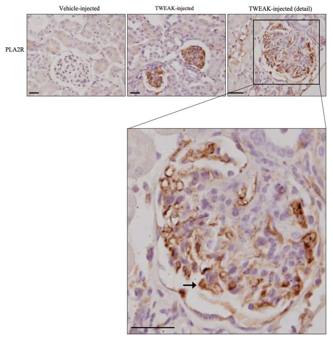 Fig. 1. TWEAK increases PLA2R expression in podocytes in vivo (Cuarental L, Valiño-Rivas L, et al., 2020).
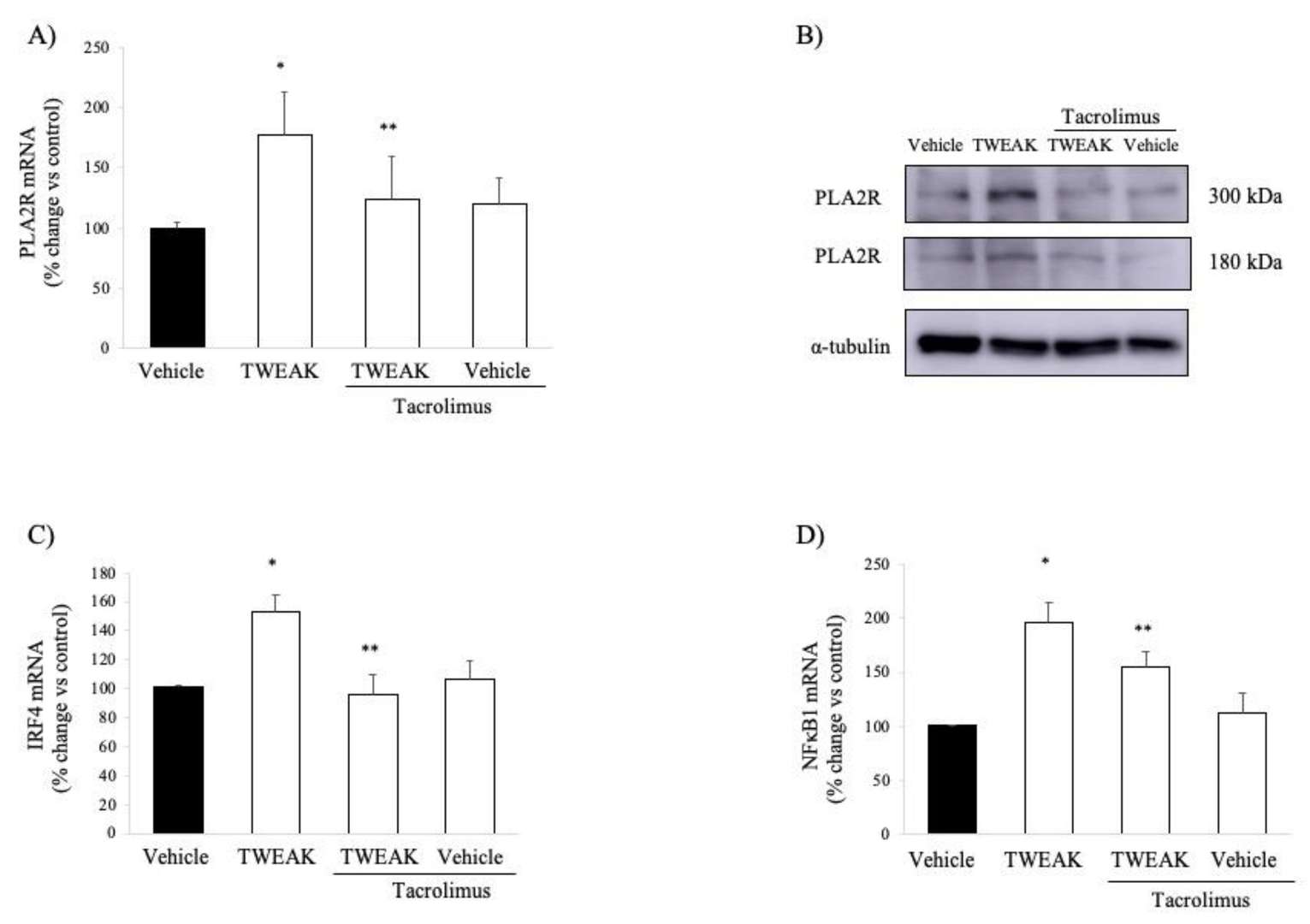 Fig. 2. TWEAK increases PLA2R expression in cultured human podocytes: impact of tacrolimus (Cuarental L, Valiño-Rivas L, et al., 2020).
Silencing of TCF7 Ameliorated HG-Triggered Injury in Podocytes
Diabetic nephropathy (DN) is a severe complication of diabetes characterized by podocyte destruction. Hyperglycemia, the most important contributor to podocyte loss, can lead DN towards diabetic nephropathy. The pathogenicity of DN involves long non-coding RNAs (lncRNAs) such as LncRNA T-cell factor 7 (TCF7) and semaphorin-3A (SEMA3A) implicated in kidney disease. But it is not clear how TCF7's effects on DN are related to SEMA3A.
Jiang's team explored TCF7's role in high glucose podocyte damage and whether it is mediated via the miR-16-5p/SEMA3A pathway. TCF7 expression significantly increased compared with glucose and mannitol groups (Fig. 3a). In order to better characterise the role of TCF7 in diabetic nephropathy, TCF7 was silenced by siRNA (si-TCF7), which knocked down TCF7 expression in high-glucose or non-hyperglycemic podocytes (Fig. 3b and c). High glucose lowered cell viability (Fig. 3d) and resulted in greater apoptosis (Fig. 3e and f) by enhancing Bax and decreasing Bcl-2 (Fig. 3g and h). It also upregulated ROS (Fig. 3i), MDA (Fig. 3j), IL-1 (Fig. 3m), TNF- (Fig. 3n), IL-6 (Fig. 3o) and decreased SOD (Fig. 3k) and CAT (Fig. 3l). TCF7 silencing blocked these podocyte-affecting high glucose levels (Fig. 3d–o).
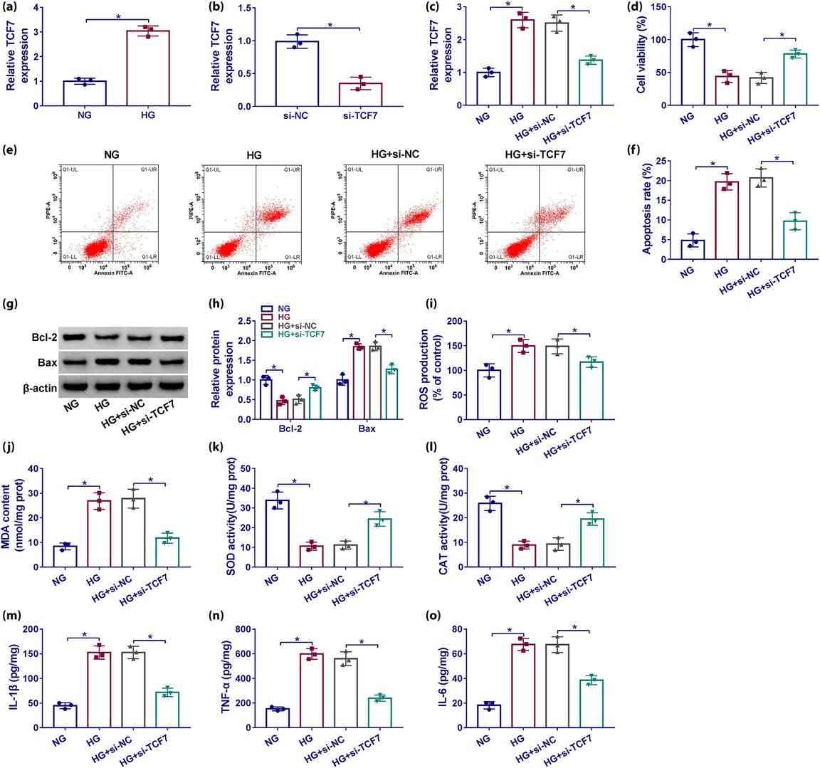 Fig. 3. TCF7 depletion protected podocytes from cytotoxicity induced by high glucose (Jiang Z, Qian L, et al., 2023).
Ask a Question
Write your own review
Description: The 293T cell line, originally referred as 293tsA1609neo, is a highly transfectable derivative of human embryonic kidney 293 cells, and contains the SV40 T-antigen
Description: Creative Bioarray offers a normal human Renal Proximal Convoluted Tubule Epithelial cell system. Renal proximal convoluted tubule epithelial cells are an essential research tool for understanding a variety of biological processes in the kidneys. They can also be utilized to determine renal toxicity of compounds as well as to study underlying causes and fundamental aspects of human kidney diseases.
These primary RPCTs are collected from unfractionated kidney cell preparations from normal, non-diseased human kidney tissues. The cells can be cultured and expanded in Bioarray's unique renal epithelial cell culture media formulations delivering a robust renal cell culture for use in experiments and screening assays.
Bioarray provides RPCTs cryopreserved at 500,000 cells/vial along with 50ml of RPCT plating media for each vial to establish initial culture. Purity of each lot is verified using morphology and CD13 flow cytometry – Bioarray RPCTs are qualified at >80% CD13+ upon QC release.
Each lot of primary renal cells are tested and verified negative for HIV-1, HIV-2, HTLV I & II, Hep B & C, Syphillis and CMV.
Custom donor specifications are available upon request and a number of different plate and flask formats can be customized to meet your particular research project needs.
Description: HRPTEpiC from Creative Bioarray Research Laboratories are isolated from human kidney. HRPTEpiC are cryopreserved at passage one and delivered frozen. Each vial contains >5 x 10^5 cells in 1 ml volume. HRPTEpiC are characterized by immunofluorescent method with antibodies to cytokeratin-18, -19 and vimentin. HRPTEpiC are negative for HIV-1, HBV, HCV, mycoplasma, bacteria, yeast and fungi. HRPTEpiC are guaranteed to further expand for 15 population doublings in the condition provided by Bioarray.
Description: HRMC from Creative Bioarray Research Laboratories are isolated from human renal tissue. HRMC are cryopreserved after purification and delivered frozen. Each vial contains >5 x 10^5 cells in 1 ml volume. HRMC are characterized by immunofluorescent method with antibodies to fibronectin, Thy-1, and smooth muscle actin. HRMC are negative for HIV-1, HBV, HCV, mycoplasma, bacteria, yeast and fungi. HRMC are guaranteed to further expand for 15 population doublings at the conditions specified by Creative Bioarray.
Description: Human Renal Cortical Epithelial Cells (HRCECs) provided by Creative Bioarray are isolated from the normal human kidney tissue. The cells are cryopreserved at passage 2 and delivered frozen. Each vial contains more than 0.5*10^6 viable cells after thawing. The cells are negative for HIV-1, HBV, HCV, mycoplasma, bacteria, yeast and fungi. HRCECs are guaranteed for at least 12 population doublings under the conditions provided by Creative Bioarray. Repeated freezing and thawing of cells is not recommended.

Description: Creative Bioarray's normal Human Renal Epithelial Cells, when grown in Creative Bioarray's LIRen Medium, provide an ideal low-serum culture model for the study of renal function, metabolism, nephrotoxicity or cancer research. Creative Bioarray's Renal Proximal Tubule Epithelial Cells are cryopreserved as secondary cells to ensure the highest viability and plating efficiency. Our Renal Epithelial Cells are quality tested in LIRen Medium to ensure optimal reduced-serum growth over a period of at least 15 population doublings at rates equal to or greater than serum-supplemented medium.
Cell Features:
Mixed Renal Epithelial, Renal Cortical Epithelial, and Renal Medullary Epithelial are cryopreserved as primary cells isolated from human kidney tissue and expanded in culture vessels once before cryopreservation.
Renal Proximal Tubule Epithelial are cryopreserved as secondary cells isolated from human kidney tissue and expanded in culture vessels twice before cryopreservation.
All Renal Epithelial Cell types can be grown in a 0.5% serum medium without phenol red or antimicrobials when cultured in LIRen Medium.
All Renal Epithelial Cell types are extensively tested for quality and optimal performance.
Creative Bioarray guarantees performance and quality.
