Featured Products
Hot Products
ONLINE INQUIRY
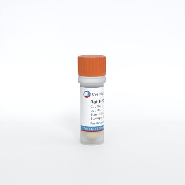
Rat Hepatic Stellate Cells
Cat.No.: CSC-C1808
Species: Rat
Source: Liver
Cell Type: Hepatic Stellate Cell
- Specification
- Q & A
- Customer Review
Cat.No.
CSC-C1808
Description
Hepatic stellate cells (HSteC) are intralobular connective tissue cells presenting myofibroblastlike or lipocyte phenotypes. They participate in the homeostasis of liver extracellular matrix, repair, regeneration, fibrosis and control retinol metabolism, storage and release. Following liver injury, HSteC transform into myofibroblast-like cells and are the major source of type I collagen in the fibrotic liver. Beyond these feature, HSteC have been implicated as regulators of hepatic microcirculation via cell contraction, and in disease states, in the pathogenesis of intrahepatic portal hypertension. Proliferation and migration of HSteC and expression of chemokines are involved in the pathogenesis of liver inflammation and fibrogenesis. HSteC possess voltageactivated calcium current, express the low affinity nerve growth factor receptor p75, and undergo apoptosis in response to nerve growth factor stimulation. Therefore, the new insight into the molecular regulation of HSteC activation will lead to therapeutic approaches in treatment of hepatic fibrosis in the future, and could lead to reduced morbidity and mortality in patients with chronic liver injury.
RHSteC are isolated from neonate day 2 rat liver tissue. RHSteC are cryopreserved immediately after purification and delivered frozen. Each vial contains >5 x 10^5 cells in 1 ml volume. RHSteC are characterized by immunofluorescent method with antibodies to desmin and ?-actin. RHSteC are negative for mycoplasma, bacteria, yeast and fungi. RHSteC are guaranteed to further expand for 5 population doublings in the conditions provided by Creative Bioarray.
RHSteC are isolated from neonate day 2 rat liver tissue. RHSteC are cryopreserved immediately after purification and delivered frozen. Each vial contains >5 x 10^5 cells in 1 ml volume. RHSteC are characterized by immunofluorescent method with antibodies to desmin and ?-actin. RHSteC are negative for mycoplasma, bacteria, yeast and fungi. RHSteC are guaranteed to further expand for 5 population doublings in the conditions provided by Creative Bioarray.
Species
Rat
Source
Liver
Recommended Medium
It is recommended to use Stellate Cell Medium for the culturing of RHSteC in vitro.
Cell Type
Hepatic Stellate Cell
Disease
Normal
Storage and Shipping
ship in dry ice; store in liquid nitrogen
Citation Guidance
If you use this products in your scientific publication, it should be cited in the publication as: Creative Bioarray cat no. If your paper has been published, please click here to submit the PubMed ID of your paper to get a coupon.
Ask a Question
Write your own review
Related Products


