ONLINE INQUIRY

Human Renal Glomerular Endothelial Cells
Cat.No.: CSC-C1559
Species: Human
Source: Kidney
Cell Type: Endothelial Cell
- Specification
- Background
- Scientific Data
- Q & A
- Customer Review
HRGEC are isolated from human kidneys. HRGEC are cryopreserved after purification and delivered frozen. Each vial contains >5 x 10^5 cells in 1 ml volume. HRGEC are characterized by immunofluorescent method with antibodies to vWF/Factor VIII and CD31 (P-CAM), and by the formation of microtublar structure in vitro. HRGEC are negative for HIV-1, HBV, HCV, mycoplasma, bacteria, yeast and fungi. HRGEC are guaranteed to expand beyond 15 population doublings at the conditions specified by Creative Bioarray.
The glomerulus, the kidney's central filter, consists of four cell populations: parietal epithelial cells, glomerular endothelial cells (GEnC), visceral epithelial cells (podocytes), and mesangial cells. HRGECs have a unique morphology of tiny transcellular pores, or fenestrations, that extend from 20 to 50 per cent of the endothelial surface. As part of the glomerular microcirculation, HRGECs filter blood. They contribute to the glomerular filtration barrier, along with the glomerular basement membrane and podocytes. This membrane blocks large proteins and blood cells from the filtrate while letting small molecules such as glucose, amino acids, and urea pass through. HRGECs also produce essential bioactive molecules and ramp up this activity under pro-inflammatory and pro-thrombotic conditions. Damage to HRGECs significantly impacts the progression and repair of glomerular diseases.
HRGECs are a useful diagnostic resource in the study of glomerular disorders, including diabetic nephropathy, hemolytic uremic syndrome and pre-eclampsia, where damage is linked to progression of disease. Moreover, given advancements in regenerative medicine, HRGECs could also be used as a cell source to treat glomerular damage.
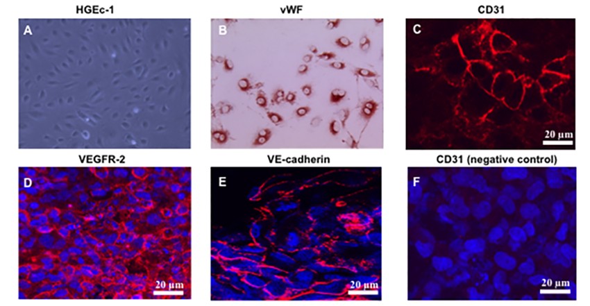 Fig. 1. Characterization of the human glomerular endothelial cell line. Immunofluorescence staining of cultured HGEc-1 for (C) CD31, (D) VEGFR-2, (E) VE-cadherin, all in red color, and (F) human podocytes incubated with the anti- CD31 antibody (Das JR, Gutkind JS, et al., 2016).
Establishment of a Microfluidic Glomerular Model Using Human Renal Glomerular Endothelial Cells
Traditional methods for drug nephrotoxicity assessment, such as animal models and static in vitro cultures, often fall short in replicating human responses. To address these limitations, microfluidic technology offers a promising alternative by recreating the dynamic human organ environment with precise control over cell microenvironments.
Qing's team employed a CellASICONIX2 Microfluidic Platform and M04S microfluidic plate for culturing human renal glomerular endothelial cells (HRGECs). Cells were prepared and introduced into the microfluidic plate, which featured multiple inlets and outlets, allowing precise control over the cell environment. After loading, cells were incubated at 37°C with 5% CO2. The microfluidic setup maintained a continuous nutrient supply through gravity-driven medium flow, facilitating cell growth in an environment that closely mimics physiological conditions (Fig. 1A and B). The growth state of the cells in the culture chamber was observed at different times, as shown in Figure 1C. The cells began to grow adherently at approximately day 1, and their morphology varied between round, triangular, fusiform, and irregular. At day 2, the confluence ratio of the cells was greater than 80%. As the incubation time increased, the cells were able to proliferate normally to form a dense cell layer at day 3. The results of live/dead cell staining showed that a well-defined confluent cell monolayer was formed, HRGECs proliferated well in cell chamber (Fig. 1D).
 Fig. 1. Design of a CellASIC-based microfluidic platform to culture glomerular endothelial cells (Qin W, Yang Z, et al., 2022).
IL-6 Increases Human Renal Glomerular Endothelial Cells Permeability
IgA nephropathy (IgAN) is a common chronic kidney disease that leads to end-stage renal failure. The pathogenesis of IgAN remains unclear, though damage to glomerular endothelial cells is known to contribute to disease progression. An inflammatory cytokine called interleukin 6 (IL-6) is thought to be involved, though its specific role in endothelial cell damage is not clear. Yang's team aims to elucidate the impact of IL-6 on glomerular endothelial cell permeability, by studying an IgAN rat model as well as cultured human renal glomerular endothelial cells (HRGECs).
They used the CCK-8 assay to see whether IL-6 influenced HRGEC permeability. Because it is unclear what IL-6's signaling pathway (classical vs trans-signaling) is, two experimental groups were created: IL-6-none (0, 50, 75, 100, 125, 150 ng/mL) and IL-6 + IL-6 receptor (IL-6R) with increased IL-6. HRGECs were treated for 24 and 48 hours, and proliferation was dramatically reduced at IL-6 levels 125 ng/mL at 48 hours (Fig. 2a). IL-6 plus 15-fold IL-6R resulted in strong inhibition at IL-6 concentrations 75 ng/mL (Fig. 2b), suggesting that IL-6 predominantly functions via the trans-signaling pathway. For further HRGEC stimulation, they then used 100 ng/mL IL-6 and 1500 ng/mL IL-6R and split the samples into normal, IL-6 and IL-6/IL-6R groups. They measured TEER using IL-6 or IL-6/IL-6R, and observed significant decreases in TEER over 24 and 48 hours, while IL-6/IL-6R had a greater impact over 48 hours (Fig. 2c). FITC-dextran tests demonstrated increased permeability in both treated groups, and especially in the IL-6/IL-6R group (Fig. 2d).
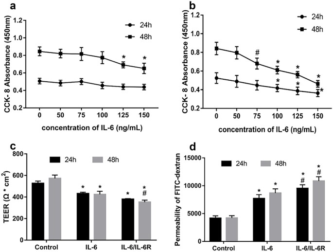 Fig. 2. Effects of IL-6 on HRGEC cell viability and permeability (Yang Y, Fu H, et al., 2022).
Ask a Question
Write your own review
Description: The 293T cell line, originally referred as 293tsA1609neo, is a highly transfectable derivative of human embryonic kidney 293 cells, and contains the SV40 T-antigen
Description: Creative Bioarray offers a normal human Renal Proximal Convoluted Tubule Epithelial cell system. Renal proximal convoluted tubule epithelial cells are an essential research tool for understanding a variety of biological processes in the kidneys. They can also be utilized to determine renal toxicity of compounds as well as to study underlying causes and fundamental aspects of human kidney diseases.
These primary RPCTs are collected from unfractionated kidney cell preparations from normal, non-diseased human kidney tissues. The cells can be cultured and expanded in Bioarray's unique renal epithelial cell culture media formulations delivering a robust renal cell culture for use in experiments and screening assays.
Bioarray provides RPCTs cryopreserved at 500,000 cells/vial along with 50ml of RPCT plating media for each vial to establish initial culture. Purity of each lot is verified using morphology and CD13 flow cytometry – Bioarray RPCTs are qualified at >80% CD13+ upon QC release.
Each lot of primary renal cells are tested and verified negative for HIV-1, HIV-2, HTLV I & II, Hep B & C, Syphillis and CMV.
Custom donor specifications are available upon request and a number of different plate and flask formats can be customized to meet your particular research project needs.
Description: HRPTEpiC from Creative Bioarray Research Laboratories are isolated from human kidney. HRPTEpiC are cryopreserved at passage one and delivered frozen. Each vial contains >5 x 10^5 cells in 1 ml volume. HRPTEpiC are characterized by immunofluorescent method with antibodies to cytokeratin-18, -19 and vimentin. HRPTEpiC are negative for HIV-1, HBV, HCV, mycoplasma, bacteria, yeast and fungi. HRPTEpiC are guaranteed to further expand for 15 population doublings in the condition provided by Bioarray.
Description: HRMC from Creative Bioarray Research Laboratories are isolated from human renal tissue. HRMC are cryopreserved after purification and delivered frozen. Each vial contains >5 x 10^5 cells in 1 ml volume. HRMC are characterized by immunofluorescent method with antibodies to fibronectin, Thy-1, and smooth muscle actin. HRMC are negative for HIV-1, HBV, HCV, mycoplasma, bacteria, yeast and fungi. HRMC are guaranteed to further expand for 15 population doublings at the conditions specified by Creative Bioarray.
Description: Human Renal Cortical Epithelial Cells (HRCECs) provided by Creative Bioarray are isolated from the normal human kidney tissue. The cells are cryopreserved at passage 2 and delivered frozen. Each vial contains more than 0.5*10^6 viable cells after thawing. The cells are negative for HIV-1, HBV, HCV, mycoplasma, bacteria, yeast and fungi. HRCECs are guaranteed for at least 12 population doublings under the conditions provided by Creative Bioarray. Repeated freezing and thawing of cells is not recommended.

Description: Creative Bioarray's normal Human Renal Epithelial Cells, when grown in Creative Bioarray's LIRen Medium, provide an ideal low-serum culture model for the study of renal function, metabolism, nephrotoxicity or cancer research. Creative Bioarray's Renal Proximal Tubule Epithelial Cells are cryopreserved as secondary cells to ensure the highest viability and plating efficiency. Our Renal Epithelial Cells are quality tested in LIRen Medium to ensure optimal reduced-serum growth over a period of at least 15 population doublings at rates equal to or greater than serum-supplemented medium.
Cell Features:
Mixed Renal Epithelial, Renal Cortical Epithelial, and Renal Medullary Epithelial are cryopreserved as primary cells isolated from human kidney tissue and expanded in culture vessels once before cryopreservation.
Renal Proximal Tubule Epithelial are cryopreserved as secondary cells isolated from human kidney tissue and expanded in culture vessels twice before cryopreservation.
All Renal Epithelial Cell types can be grown in a 0.5% serum medium without phenol red or antimicrobials when cultured in LIRen Medium.
All Renal Epithelial Cell types are extensively tested for quality and optimal performance.
Creative Bioarray guarantees performance and quality.
