ONLINE INQUIRY

Human Splenic Endothelial Cells (HSEC)
Cat.No.: CSC-7710W
Species: Human
Source: Spleen
Cell Type: Endothelial Cell
- Specification
- Background
- Scientific Data
- Publications
- Q & A
- Customer Review
The spleen operates as a vital lymphatic organ in humans by performing essential roles such as immune surveillance and filtering blood. The complex internal vascular structure of the spleen consists of endothelial cells where human splenic endothelial cells (HSECs) serve as a vital part of the splenic vascular endothelium. HSECs serve as the lining for the splenic vascular system by covering the splenic sinuses and trabecular veins to create a selective barrier that enables precise cellular and molecular exchange between blood and splenic tissue. The human splenic endothelial cells show standard endothelial morphology with polygonal or spindle-shaped forms. HSECs display enhanced permeability compared to regular capillary endothelial cells because they lack tight intercellular junctions and full basement membrane structure. Instead, they feature a developed fenestration structure, which is an essential conduit for substance exchange in the splenic microenvironment, allowing the free passage of specific cells, nutrients, and metabolic products.
The splenic immune response relies heavily on HSECs for its immunological function. Their ability to control leukocyte extravasation and immune cell activation establishes them as a perfect model to study splenic immune functions alongside immune tolerance mechanisms. Meanwhile, the high permeability of HSECs makes them ideal for investigating how substances move between blood and splenic tissue.
CD14dim/CD16 Monocytes Induce VWF:Ag Release from Splenic ECs, In Vitro
Desmopressin acetate (DDAVP) is an established treatment for mild haemophilia A and certain von Willebrand diseases, known for its rapid induction of von Willebrand factor (VWF) and factor VIII release. Research suggests that some peripheral blood mononuclear cells, potentially mediated by the spleen, express AVPR2 and might influence endothelial cell response to DDAVP.
Hypothesizing that DDAVP-responsive cells are located in the spleen, Salarilak et al. conducted an in vitro study to examine DDAVP's effect on human splenic endothelial cells (HSECs) in the presence of two monocyte subtypes: classical (CD14/CD16 ++−) and non-classical (CD14dim/CD16). Neonatal and adult HSECs co-produce factor VIII and VWF, with 0.2 mU/cm² FVIII:C and 3 mU/cm² VWF:Ag detected in the 48-hour supernatant up to passage 4. Production declines, ceasing FVIII:C detection by passage 6. In the study, HSECs were treated with DDAVP (100 ng/ml) for 1 hour in monoculture or co-culture with PBMCs or monocytes. VWF:Ag and P-Selectin:Ag levels in supernatants were measured. DDAVP alone did not increase VWF and P-selectin release unless CD14dim/CD16 monocytes were present, where significantly more VWF:Ag was found compared to untreated cells. DDAVP's effect on HSECs may involve a signal from CD14dim/CD16 monocytes, with platelet-activating factor (PAF) as a possible transmitter. They tested this by treating HSECs with various PAF concentrations (12 ng/ml, 1 μg/ml, and 40 μg/ml). Figure 1 shows that while PMA significantly increased VWF:Ag release, neither PAF nor DDAVP had this effect.
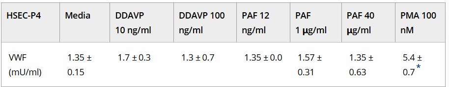 Fig. 1. VWF:Ag released from treated human splenic endothelial cells (Salarilak L, Pirdel Z, et al., 2022).
Vwf Expression is Increased and Gene Expression Profiles are Changed in HSEC at Low Temperature
von Willebrand factor (vWF) is essential for hemostasis, forming bridges with platelets after vascular injury. While previous studies have shown that hypothermia can enhance vWF production and lead to platelet activation in the spleen, the precise mechanisms involved remain unclear. Hypothermia, though sometimes used as a neuroprotective strategy, can cause coagulation disorders by affecting platelet function. Horioka's team aim to elucidate gene expression changes induced by cold stimulation to understand the molecular basis for vWF expression during hypothermia.
They first evaluated the impact of low temperature on vWF expression in HSEC, finding that it upregulated vWF mRNA and protein levels, as shown by real-time RT-PCR analysis (Fig. 2A and B). This suggests low temperature enhances vWF expression in HSEC. DNA microarray analyses were conducted to identify temperature-induced gene expression changes in HSECs. Fig. 3A shows that of 24,460 genes analyzed, those with an absolute fold change >2.0 were chosen for further study. Comparison between control and cold-treated groups revealed changes in 2,440 HSEC genes. Pathway analysis highlighted that the top ten pathways affected related to blood vessel morphogenesis, angiogenesis, and inflammation (Fig. 3B). FOS and EGR1, key transcription factors, regulate these pathways. Genes with an absolute fold change >8.0 included 6 upregulated (such as FOS and EGR1) and 71 downregulated genes.
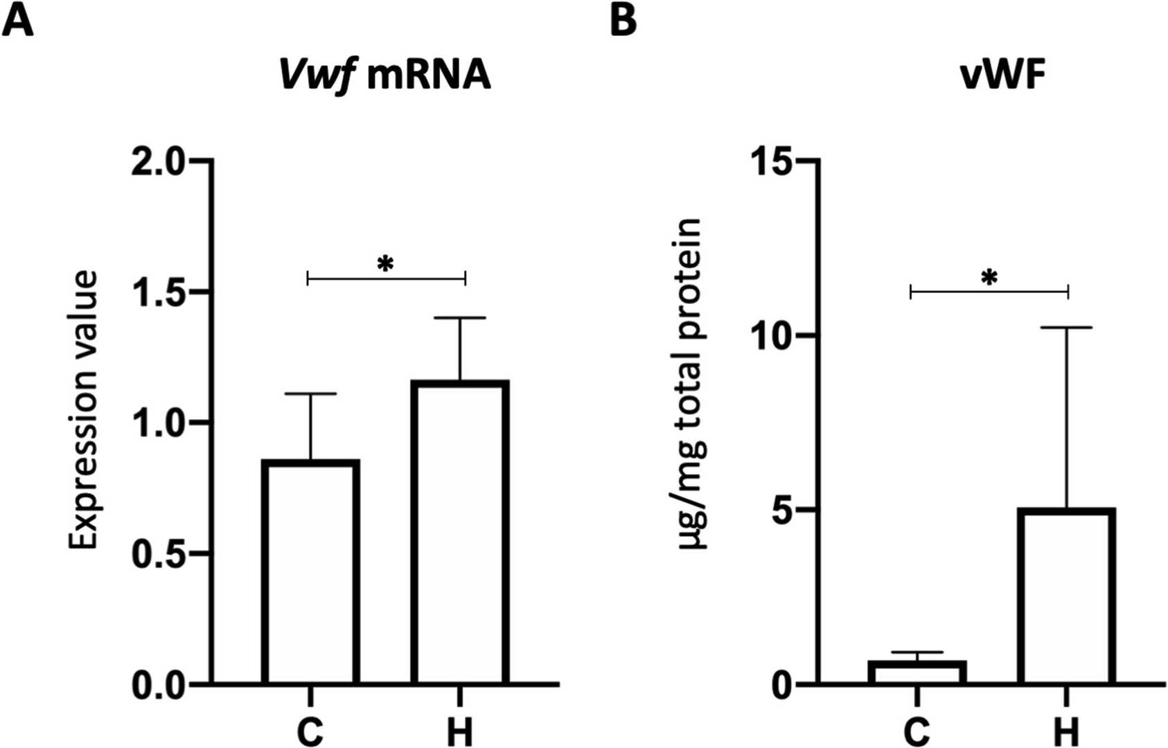 Fig. 2. vWF expression in HSEC cultured at a low temperature (Horioka K, Tanaka H, et al., 2020).
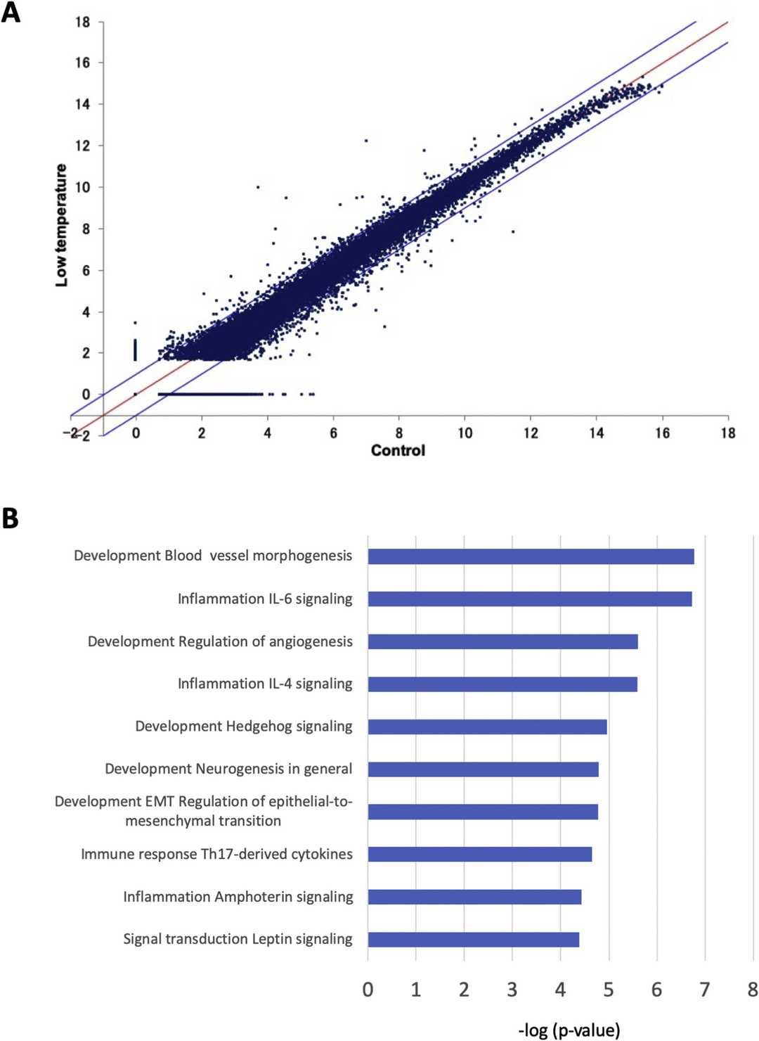 Fig. 3. cDNA microarray in HSEC (Horioka K, Tanaka H, et al., 2020).
Ask a Question
Write your own review
Description: HSF from Creative Bioarray are isolated from human spleen tissue. HSF are cryopreserved at primary culture and delivered frozen. Each vial contains >5 x 10^5 cells in 1 ml volume. HSF are characterized by their spindle morphology and immunofluorescent method with antibody to fibronectin. HSF are negative for HIV-1, HBV, HCV, mycoplasma, bacteria, yeast and fungi. HSF are guaranteed to further expand for 15 population doublings at the condition provided byCreative Bioarray.
Description: Human Spleen Fibroblasts are isolated from normal human spleen tissue.
Description: Human Spleen Microvascular Endothelial Cells are isolated from human spleen tissue.
Description: Human Spleen Epithelial Cells are isolated from normal human spleen tissue.
