ONLINE INQUIRY

Human Small Intestinal Microvascular Endothelial Cells
Cat.No.: CSC-C8618W
Species: Human
Source: Small Intestine; Intestine
Cell Type: Endothelial Cell; Microvascular Cell
- Specification
- Background
- Scientific Data
- Q & A
- Customer Review
Human small intestinal microvascular endothelial cells are isolated from the microvascular wall of human small intestinal tissue. Small intestine between gastric pylorus and cecum is the largest organ of the digestive system, and its primary place of defecation and absorption. These cells are normally cobblestone-shaped, and feature prominent, dark-colored nuclei under the light microscope. They can be detected by the surface markers CD31 and von Willebrand factor (vWF). These cells are one of the primary cell types of the vessel walls that innervate the blood vessels, tightly packed so that a barrier keeps the artery's walls open so that blood-borne molecules don't get into the gut randomly. They also keep material from gastrointestinal tissues out of the bloodstream, thus mediating material flow between blood and gastrointestinal tissues. During inflammation, microvascular endothelial cells can capture and attach to on-circulating immune cells, which regulate inflammation.
In organ chip construction, co-culturing small intestinal microvascular endothelial cells with intestinal epithelial cells on the chip can simulate various physiological functions of the small intestine, such as the transport, absorption, and metabolism of nutrients. The fluid flow and peristalsis-like mechanical movements within the chip also allow for the study of dynamic intestinal processes. For instance, this organ chip model can be used to deeply investigate mechanisms and pathological processes related to small intestinal diseases, such as inflammatory bowel disease and intestinal obstruction.
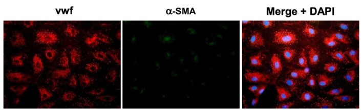 Fig. 1. Immunofluorescence images of human intestinal microvascular endothelial cells labeling with von Willebrand factor (VWF), a-smooth muscle actin (a-SMA) and DAPI (Mintet E, Rannou E, et al., 2015).
Development the Adult Duodenum Intestine-Chip Use Human Small Intestinal Microvascular Endothelial Cells
Orally administered drugs face bioavailability challenges mainly due to intestinal drug-drug interactions and enzyme activities. Traditional models, such as Caco-2, lack three-dimensional cytoarchitecture and accurate enzyme expression. Therefore, Kasendra's team combined intestinal organoids and Organs-on-Chips technology to develop a Duodenum Intestine-Chip. In summary, organoid cultures were established from biopsied crypts of healthy adults (Fig. 1A; top), and fragments were seeded on ECM-coated PDMS membranes in the chips (Fig. 1B; 3: epithelial tissue). Human small intestinal microvascular endothelial cells (HIMECs) (Fig. 1A; bottom) populated the PDMS membrane in the vascular channel (Fig. 1B; 4: endothelial cells). The Duodenum Intestine-Chip was continuously perfused with fresh medium, and cyclic mechanical strain (10% strain, 0.2 Hz) was applied to mimic intestinal peristalsis.
They evaluated the effects of mechanical stimulation on the phenotype of the intestinal cells using immunofluorescence and SEM for microvilli identification. Exposure to flow for 72 hours induced epithelial polarization and microvilli formation (Fig. 1 and Fig. 2), aligning with previous Caco-2 cell findings. Static cultures formed flat squamous cells with poor junctions (Fig. 1 and Fig. 2A) and sparse microvilli (Fig. 1 and Fig. 2B), while flow conditions yielded well-polarized cells with distinct junctions and dense microvilli. Continuous flow was crucial for epithelial maturation, and extended exposure to flow and strain led to 'villi-like structures' by day 6 (Fig. 1C). Confocal analysis showed confluent monolayers with defined tight and adherent junctions (Fig. 1D). These conditions improved intestinal permeability over time, indicated by low dextran permeability (Papp) in chips from different individuals (Fig. 1E). Overall, this data indicates that the human adult Duodenum Intestine-Chip supports the formation of a functional barrier with in vivo relevant cytoarchitecture, cell-cell interactions, and permeability parameters.
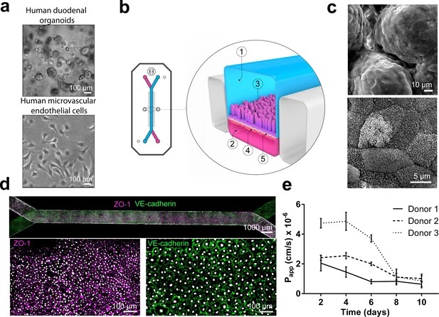 Fig. 1. Duodenum Intestine-Chip: a microengineered model of the human duodenum (Kasendra M, Luc R, et al., 2020).
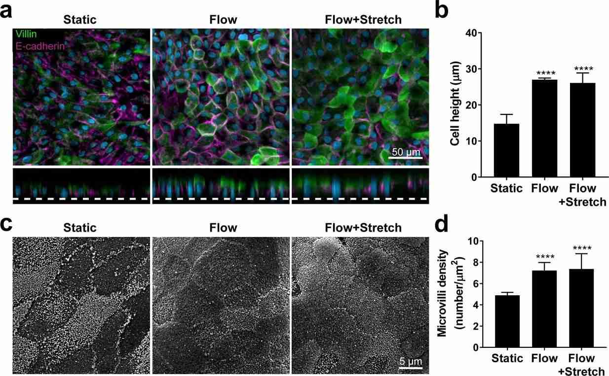 Fig. 2. Flow-induced increase in primary human small intestinal epithelial cells height and microvilli formation (Kasendra M, Luc R, et al., 2020).
Neonatal-intestine-on-a-chip Models Human Small Intestinal Architecture
Necrotizing enterocolitis (NEC) is a severe intestinal disease in premature infants, characterized by excessive inflammation, bacterial imbalance, and epithelial damage leading to a disrupted gut barrier. Lanik et al. developed a Neonatal-Intestine-on-a-Chip to model NEC, which utilizes a combination of premature infant small intestinal enteroids cocultured with human small intestinal microvascular endothelial cells and patient-derived microbiota to recreate critical features of premature gut pathophysiology, including microbial dysbiosis.
To establish baseline cellular growth, they tracked the development of intestinal stem cells from enteroids (day 0) to neonatal epithelium (day 8) using brightfield microscopy (Fig. 3A). On day 0, enteroids are distinct organoids. By day 1, they were dissociated and seeded into a Matrigel-coated microfluidic device, forming a confluent monolayer by day 3. Furrows appeared by day 6, progressing to a 3D architecture with villus-like structures by day 8 (Fig. 3A). To assess architectural similarity to the human small intestine, they immunostained cells for villi markers: villin (green) and FABP1 (white), with counterstaining by phalloidin (red) and Hoechst 33342 (blue; Fig. 3B). The resulting structure resembles human intestinal villi with microvilli-lined enterocytes. They examined polarity within the Neonatal-Intestine-on-a-Chip using ZO-1 (red) for apical junctions and β-catenin (green) for basolateral junctions (Fig. 3C). Confocal microscopy showed ZO-1 near the lumen and β-catenin near the semipermeable membrane, indicating key neonatal intestinal features such as 3D architecture, cell adhesion, and polarity.
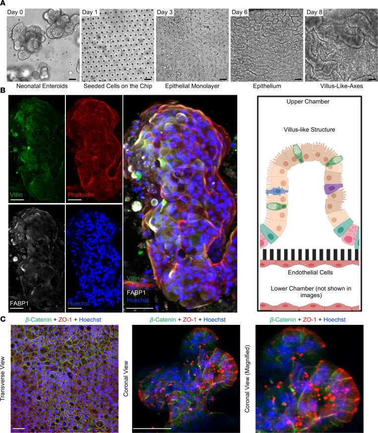 Fig. 3. Development of the Neonatal-Intestine-on-a-Chip microfluidic model (Lanik W E, Luke C J, et al., 2023).
Ask a Question
Write your own review
Description: Smooth muscle contraction is the fundamental event in gastrointestinal motion. Inflammation of the human intestine causes thickening of the smooth muscle layers which results from the increases in the smooth muscle-specific actins. The increased smooth muscle actins may affect force production and further demonstrate the plasticity of smooth muscle in the inflamed intestine. Human intestinal smooth muscle cells respond to IL-1-beta and TNF-alpha stimulation by releasing IL-6, which might significantly contribute to the overall systemic inflammatory response. Knowledge of molecular mechanism that underlies the control of colorectal smooth muscle tone is essential to advance of understanding of pathophysiology of the abnormality. The availability of human rectal smooth muscle cells in culture will considerably enhance our ability to study the contractile, proliferative and connective tissue responses of the smooth muscle of the human colorectal disorders.HRSMC from Bioarray Research Laboratories are isolated from the human rectum. HRSMC are cryopreserved at primary or passage one culture and delivered frozen. Each vial contains >5 x 10^5 cells in 1 ml volume. HRSMC are characterized by immunofluorescent method with antibodies to Α-smooth muscle actin and desmin. HRSMC are negative for HIV-1, HBV, HCV, mycoplasma, bacteria, yeast and fungi. HRSMC are guaranteed to further expand for 15 population doublings in the condition provided by Bioarray Research Laboratories.
Description: Smooth muscle contraction is the fundamental event in gastrointestinal motion. Although many of the biochemical mechanisms underlying the excitation-contraction coupling are not yet defined, it is known that cytosolic Ca2+ is the essential component in the coupling phenomenon. Inflammation of the human intestine causes thickening of the smooth muscle layers which results from the increases in the smooth muscle-specific actins. The increased smooth muscle actins may affect force production and further demonstrate the plasticity of smooth muscle in the inflamed intestine. Studies also show that human intestinal smooth muscle cells respond to IL-1beta and TNF-alpha stimulation by releasing IL-6, which might significantly contribute to the overall systemic inflammatory response. The availability of human intestinal smooth muscle cells in culture will considerably enhance our ability to study the contractile, proliferative and connective tissue responses of the smooth muscle of the human gastrointestinal tract.HISMC from Bioarray Research Laboratories are isolated from human intestine. HISMC are cryopreserved at passage one culture and delivered frozen. Each vial contains >5 x 10^5 cells in 1 ml volume. HISMC are characterized by immunofluorescent method with antibodies to Α-smooth muscle actin and desmin. HISMC are negative for HIV-1, HBV, HCV, mycoplasma, bacteria, yeast and fungi. HISMC are guaranteed to further expand for 15 population doublings in the condition provided by Bioarray Research Laboratories.
Description: Endothelial cells lining the microvasculature are known to play a critical "gatekeeper" role in the inflammatory process through their ability to recruit circulating immune cells into tissues and foci of inflammation. Studies show that intestinal microvascular endothelial cells (IMEC) exhibit a strong immune response to LPS challenge and play a critical regulatory role in gut inflammation. Pharmacological inhibition of NOS in activated HIMEC resulted in a significant increase in leukocytes binding. Gene expression profile study reveals that intestinal endothelial cells express biotinidase, which is involved in biotin recycling. The in vitro culture of HIMEC enabled scientists to perform systematic analyses of the cytokine profiles with regard to mRNA expression and protein secretion, and to compare such data with cytokine profiles concomitantly displayed by other endothelial cells.HIMEC from Bioarray Research Laboratories are isolated from human intestinal tissue. HIMEC are cryopreserved at passage one and delivered frozen. Each vial contains >5 x 10^5 cells in 1 ml volume. HIMEC are characterized by immunofluorescent method with antibodies to vWF/Factor VIII and CD31 (P-CAM) and by uptake of DiI-Ac-LDL. HIMEC are negative for HIV-1, HBV, HCV, mycoplasma, bacteria, yeast and fungi. HIMEC are guaranteed to further culture in the conditions provided by Bioarray Research Laboratories.
Description: Fibroblasts are mesenchymal cells derived from the embryonic mesoderm. They have been extensively used for a wide range of cellular and molecular studies. This is mainly because they are one of easiest types of cells to grow in culture, and their durability makes them amenable to a wide variety of manipulations ranging from studies employing gene transfection to microinjection. There is good evidence that fibroblasts in different parts of the body are intrinsically different. Fibroblasts secrete a non-rigid extracellular matrix that is rich in type I and/or type III collagen. They are responsible for much of the synthesis of extracellular matrix in connective tissues and play major roles in wound healing. Many diseases are associated with fibroblasts, either because fibroblasts are implicated in their etiology or because of the fibrosis that accompanies damage to other cell types in tissues. For example, the development of bowl stenosis in Crohn's disease patients is caused by extreme fibroblast proliferation and extracellular matrix expansion.
HIF are isolated from human intestinal tissue. HIF are cryopreserved at passage one and delivered frozen. Each vial contains >5 x 10^5 cells in 1 ml volume. HIF are characterized by their spindle morphology and immunofluorescent method with antibody to fibronectin. HIF are negative for HIV-1, HBV, HCV, mycoplasma, bacteria, yeast and fungi.
Description: Smooth muscle is responsible for the contractility of hollow organs, such as blood vessels, the gastrointestinal tract, the bladder, and the uterus. Its structure differs greatly from that of skeletal muscle. The human stomach contains three layers of muscle in its walls, the outer longitudinal, the middle circular and the inner oblique and visceral smooth muscle cells makes up all three layers along the entire organ. Smooth muscle contraction is critical to peristalsis in the human stomach and the contraction may be mediated by activation of phospholipase through two distinct mechanisms (increased intracellular Ca2+ and G protein activation) and activating PKCepsilon-dependent mechanisms. In vitro study also shows that gastric smooth muscle cells express ET and eNOS and both calcium and sodium may be involved as current carriers in the generation of the plateau potential.HGSMC from Bioarray Research Laboratories are isolated from the human stomach. HGSMC are cryopreserved at secondary culture and delivered frozen. Each vial contains >5 x 10^5 cells in 1 ml volume. HGSMC are characterized by immunofluorescent method with antibodies to Α-smooth muscle actin and desmin. HGSMC are negative for HIV-1, HBV, HCV, mycoplasma, bacteria, yeast and fungi. HGSMC are guaranteed to further expand for 15 population doublings at the condition provided by Bioarray Research Laboratories.
Description: Primary Human Intestinal Microvascular Endothelial Cells were initiated by elutriation from dissociated normal human small intestine tissue.
These cells were originated using Complete Serum-Free Medium Kit With SuperFuel™, are available at <12 Cumulative Population Doublings (CPD) in vitro [Passage 3] and were cryopreserved in aliquots of ~1.5 X 10^6 cells. This vial will initiate a Passage 4 cell culture in a 75cm2 flask.
