Featured Products
Our Promise to You
Guaranteed product quality, expert customer support

ONLINE INQUIRY
HSC-4
Cat.No.: CSC-C6352J
Species: Human
Morphology: epithelial-like
Culture Properties: Adherent cells
- Specification
- Background
- Scientific Data
- Q & A
- Customer Review
Store in liquid nitrogen.
The HSC-4 cell line is a human tongue squamous cell carcinoma (SCC) cell line established by a patient with oral cancer. This cell line was derived to facilitate research focused on the molecular and biological features of tumors that arise in the oral cavity, particularly in the tongue, which is a common site for squamous cell carcinoma. Understanding the behavior and characteristics of HSC-4 is essential for developing targeted therapies and improving treatment outcomes for patients with this type of cancer.
The cells exhibit typical features associated with malignancy, such as altered growth patterns, invasion capabilities, and resistance to standard therapeutic approaches. Researchers utilize the HSC-4 cell line to explore various aspects of cancer biology, including cellular signaling pathways, gene expression patterns, and the interaction between cancer cells and the surrounding microenvironment.
In addition to its role in basic research, HSC-4 has been instrumental in preclinical studies aimed at testing new therapeutic strategies. The cell line serves as a platform for evaluating the efficacy of chemotherapeutic agents and targeted therapies, providing insights into potential resistance mechanisms that tumors may develop.
Induction of Autophagy Impairs Migration and Invasion in HSC-4 Cells
To investigate the involvement of autophagy in cell migration and invasion, serum deprivation (serum-free media) was performed to induce autophagy in HSC-4 cells. As shown in Fig. 1 A-C, the intensity of MDC-stained autophagic vacuoles, the number of MDC-positive cells, LC3 gene expression and LC3 immunofluorescence intensity were distinctly increased in serum-free medium whereas chloroquine (CQ), an inhibitor of autophagy, pre-incubation significantly reduced MDC staining, expression level of LC3 and immunofluorescence intensity of LC3 gene compared to serum-free medium. In addition, HSC-4 cells in serum-free medium displayed a significant decrease in migration percentage, invaded the area, and number of invaded cells. In contrast, the wound area distance significantly increased compared to control cells and untreated cells. In contrast, CQ pre-treatment led to significantly abolishing the effect of autophagy on the percentage of migration, the invaded area, and the invaded cells (Fig. 1 D-F, respectively). These results demonstrate that the induction of autophagy impaired the migration and invasion capacity of HSC-4 cells.
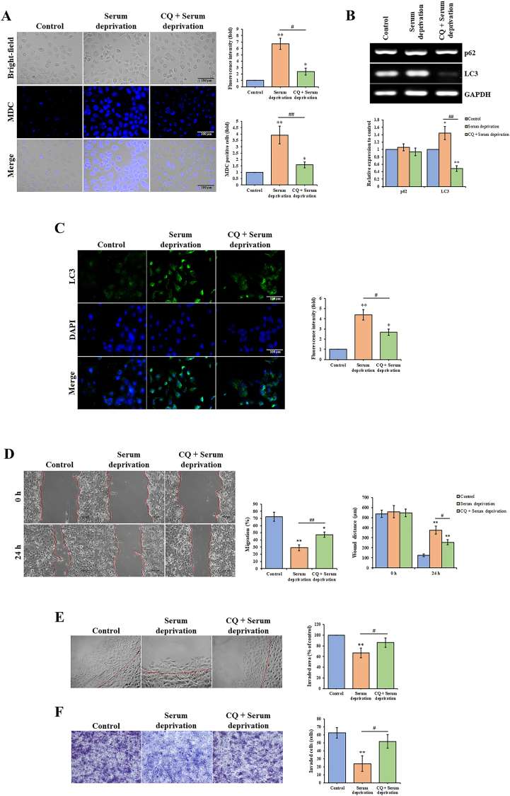 Fig. 1 Autophagy induction impairs migration and invasion in HSC-4 cells. (A) MDC staining, (B) mRNA expression of p62 and LC3 genes, and (C) LC3 immunofluorescence staining in HSC-4 cells pre-treated with 50 μM CQ for 1 h, followed by incubation in serum-free medium (serum deprivation) for 24 h. (D) Migratory ability was assessed by wound healing assay, (E and F) invasive ability was examined by the agarose spot and Transwell invasion assays in HSC-4 cells pre-treated with 50 μM CQ for 1 h, followed by incubation in serum-free medium for 24 h. (Binlateh T, et al., 2022)
Silencing of FANCD2 Increased the Bax/Bcl2 Ratio and Activated the p38 MAPK Signaling Pathway in HSC-4 Cells
Western blot analysis revealed that after the FANCD2 gene expression in HSC-4 cells was silenced by shRNA interference, the expression of the Bax and p-p38 proteins was significantly higher in the cells of the experimental group than in the control groups (P<0.05), whereas the expression of the p38 and Bcl2 proteins was significantly decreased (P<0.05; Fig. 2A and B). After radiotherapy, the same results were obtained; additionally, a significant difference was observed in the expression of these proteins before and after radiotherapy (P<0.05; Fig. 2A and B). After the nude mice with transplanted HSC-4 cells received radiotherapy, western blotting, and immunohistochemistry were performed, which showed an increase in the expression of Bax and p-p38 proteins in the tumor tissues of mice in the experimental group compared with the control group (P<0.05); in contrast, the expression of p38 and Bcl2 proteins was decreased (P<0.05; Figs. 2C and D, 3 and 4). This finding is consistent with the results of the in vitro cell culture experiments. These results suggest that the silencing of FANCD2 increases the Bax/Bcl2 ratio and activates the p38 MAPK signaling pathway in HSC-4 cells after radiotherapy.
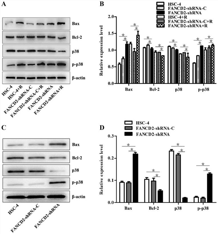 Fig. 2 (A and B) Western blot analysis was used to determine the expression of the Bax, Bcl2, p38, and p-p38 proteins in the three groups of HSC-4 cells before and after radiotherapy. (C and D) Western blot analysis was used to examine the expression of the Bax, Bcl2, p38, and p-p38 proteins in the tumors derived from transplanted HSC-4 cells after radiotherapy. (Feng HJ, et al., 2017)
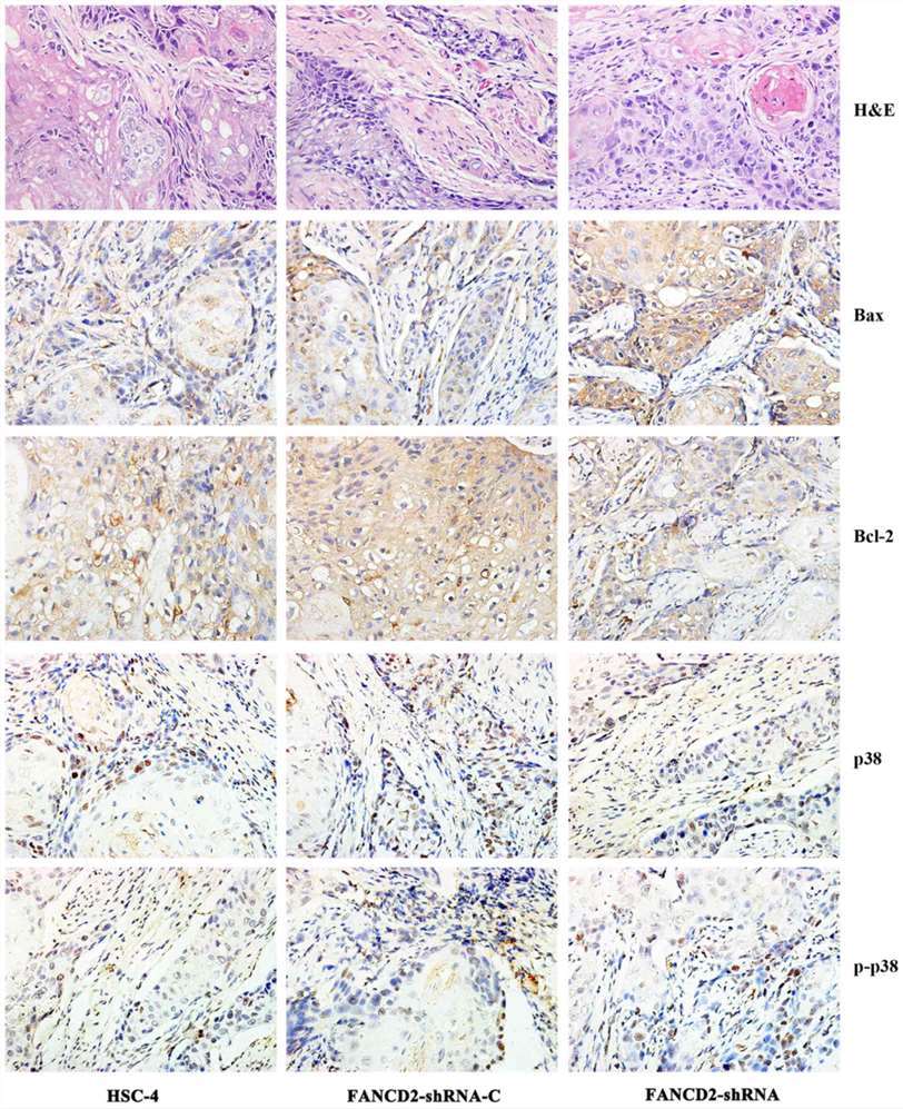 Fig. 3 H&E staining of the tumors derived from HSC-4 cells and immunohistochemical analysis of the Bax, Bcl2, p38, and p-p38 protein expression after radiotherapy. (Feng HJ, et al., 2017)
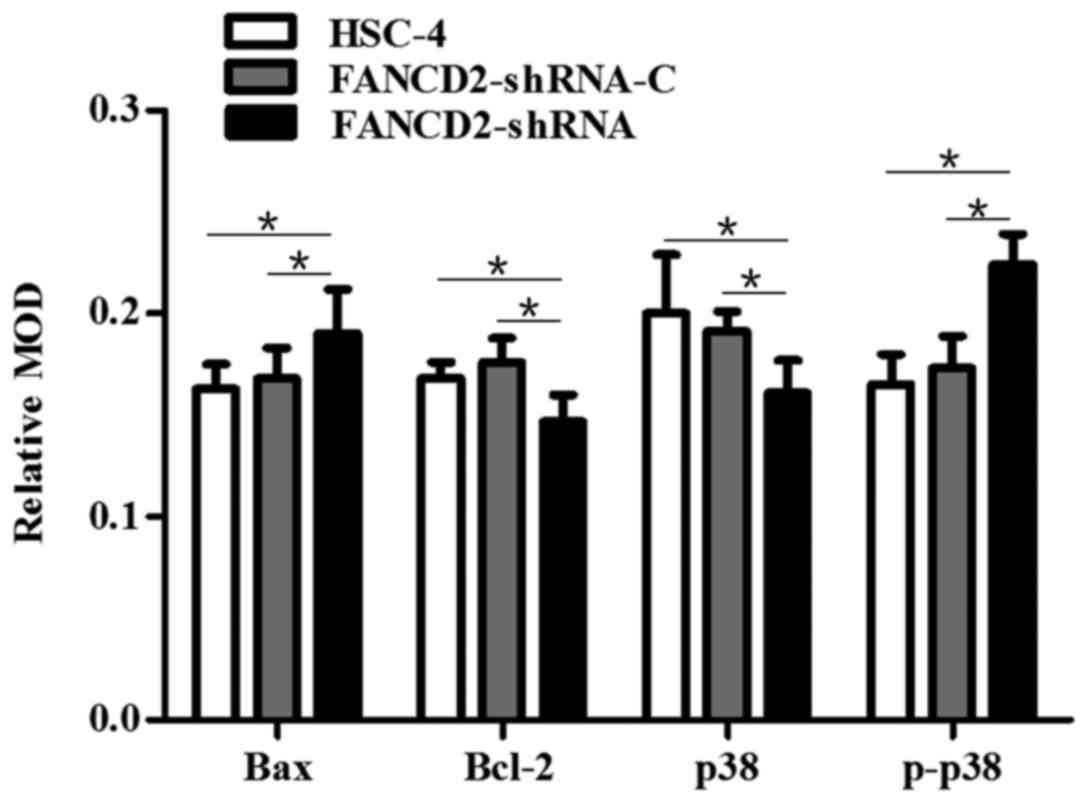 Fig. 4 Image-Pro Plus 6 software was used to quantitatively analyze the expression of the Bax, Bcl2, p38, and p-p38 proteins using immunohistochemistry (IHC) in the tumor sections from the three groups of mice after radiotherapy. (Feng HJ, et al., 2017)
High-resolution/high-mass accuracy mass spectrometers are used to identify protein phosphorylation sites due to their speed, sensitivity, selectivity, and throughput.
Ask a Question
Average Rating: 5.0 | 1 Scientist has reviewed this product
Expanded research scope
We have expanded our research scope by utilizing tumor cell products from Creative Bioarray to investigate the influence of tumor cells on the immune system.
20 Mar 2023
Ease of use
After sales services
Value for money
Write your own review
- You May Also Need

