Featured Products
Our Promise to You
Guaranteed product quality, expert customer support

ONLINE INQUIRY
HSC-2
Cat.No.: CSC-C6355J
Species: Human
Morphology: epithelial-like
Culture Properties: Adherent cells
- Specification
- Background
- Scientific Data
- Q & A
- Customer Review
Store in liquid nitrogen.
The HSC-2 cell line is a human cell line derived from oral squamous cell carcinoma (OSCC) of a 69-year-old male patient. Established to facilitate cancer research, HSC-2 provides a valuable model for investigating the characteristics and behaviors of oral cancers, which are known for their aggressive nature and complex treatment challenges.
One of the notable features of the HSC-2 cell line is its expression of HLA-A24, a specific human leukocyte antigen (HLA) class I allele. This characteristic makes HSC-2 particularly relevant for studies involving immune responses to cancer, as HLA molecules play a crucial role in presenting antigens to T cells. The study of HSC-2 can help researchers understand the interactions between the immune system and OSCC, potentially leading to the development of immunotherapeutic strategies that harness the body's immune response against tumor cells.
In addition to its immunological relevance, HSC-2 serves as a platform for preclinical testing of various treatment modalities, including chemotherapeutic agents and targeted therapies. Researchers can utilize HSC-2 to assess drug efficacy, explore mechanisms of drug resistance, and identify potential therapeutic targets.
Podocalyxin Is Crucial for HSC-2 Cell Growth
CRISPR/Cas9 plasmids targeting human podocalyxin (PODXL) were used to generate three PODXL-knock out (PODXL-KO) OSCC cell lines: HSC-2/PODXL-KO #1, #2, and #3. Before initiating the study, PODXL expression was confirmed in these cell lines. As shown in Fig. 1, anti-human PODXL mAb, PcMab-47 reacted with the HSC-2 parental cell line but did not react with any of the three HSC-2/PODXL-KO cell lines. The potential involvement of PODXL in the stimulation of in vitro OSCC cell growth. At every plated cell density (1500, 3000, and 6000 cells/well), the growth of HSC-2/PODXL-KO #1 and #2 cells was significantly lower than that of HSC-2 cells (Fig. 2). In contrast, HSC-2/PODXL-KO #3 showed significantly less growth than HSC-2 cells only when at a plated cell density of 1500 cells/well.
The role of PODXL in OSCC tumor growth in vivo was further examined by comparing the growth of four cell lines, parental HSC-2, HSC-2/PODXL-KO #1, HSC-2/PODXL-KO #2, and HSC-2/PODXL-KO #3, which were transplanted subcutaneously into nude mice. On days 7, 14, and 21 after inoculation, tumor volumes them were measured (Fig. 3). On day 7, no difference in tumor volumes was observed between parental HSC-2 and HSC-2/PODXL-KO cell lines. The tumor volume of HSC-2/PODXL-KO #1 cell lines was significantly lower than that of parental HSC-2 cell lines on day 14, although the tumor volumes for HSC-2/PODXL-KO #2 and #3 cell lines were similar to those arising from the parental cell line, HSC-2 (Fig. 3A). On day 21, tumor volumes arising from the transplantation of HSC-2/PODXL-KO #1, #2, and #3 cell lines were significantly lower than those arising from the parental HSC-2 cell lines (Fig. 3A). Subcutaneous tumors arising from parental HSC-2, HSC-2/PODXL-KO #1, HSC-2/PODXL-KO #2, and HSC-2/PODXL-KO #3 on day 21 are shown in Fig. 3B. In vivo analysis revealed that HSC-2/PODXL-KO #1 cell lines resulted in the smallest tumor growth among the three HSC-2/PODXL-KO cell lines (Fig. 3A), Taken together, our results indicate that PODXL plays an important role in the growth of tumor in oral cancers.
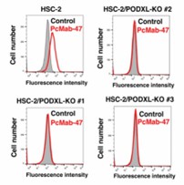 Fig. 1 Flow cytometry using HSC-2/PODXL-KO cell lines. (Itai S, et al., 2018)
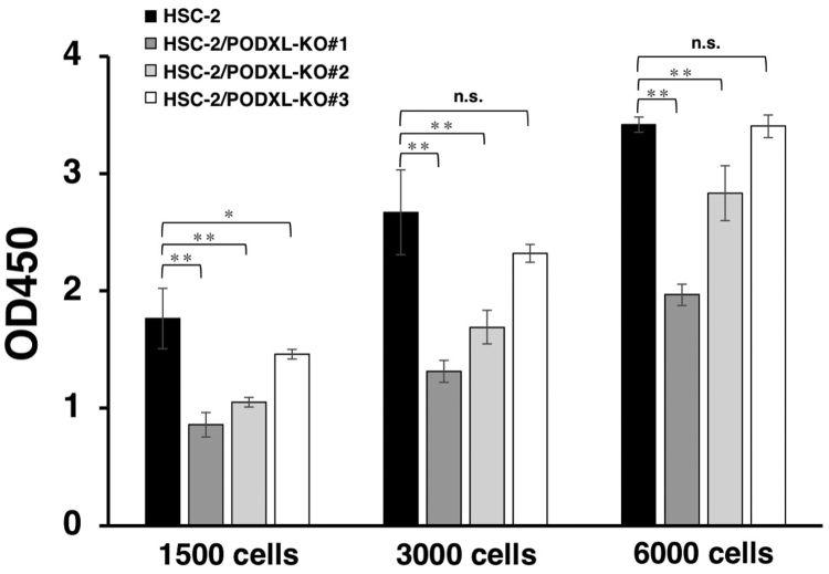 Fig. 2 In vitro functional analysis of PODXL using PODXL-KO OSCC lines. (Itai S, et al., 2018)
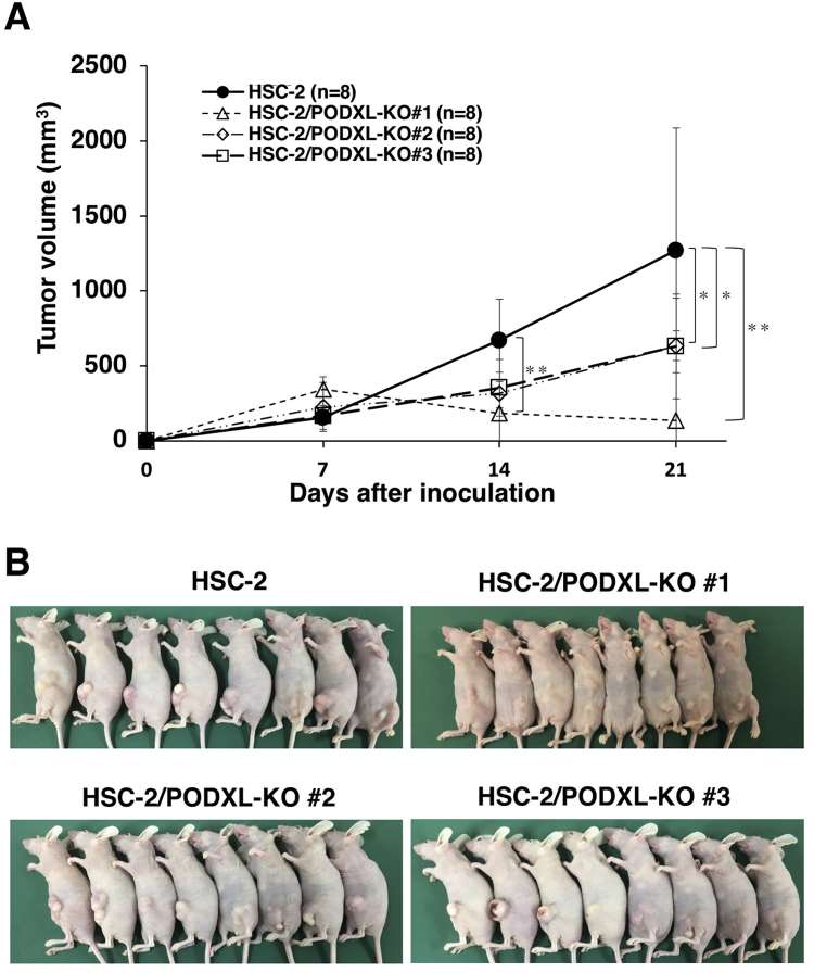 Fig. 3 In vivo functional analysis of PODXL using PODXL-KO OSCC lines. (Itai S, et al., 2018)
Antitumor Activities of H2Mab-19 in the Mouse Xenografts of Oral Cancers
H2Mab-19 possessed antitumor activity in mouse xenografts of breast cancers. Whether this activity extended to xenografts of oral cancers was also assessed. BT-474 cells expressed high levels of human epidermal growth factor receptor 2 (HER2) (Fig. 4A); HER2 levels were however low in HSC-2 cells (Fig. 4B). Nevertheless, HSC-2 is useful for the investigation of antitumor activity in vivo. Thus, HSC-2 was used for mouse xenografts of oral cancers.
HSC-2 cells were implanted subcutaneously into the flanks of nude mice. H2Mab-19 and mouse IgG were injected i.p. three times (on days 1, 6, and 14 after cell injections into treated and control mice, respectively. Tumor formation was observed in mice in both groups. In comparison to control mice, H2Mab-19-treated mice showed significantly reduced tumor development on days 6, 10, 14, 17, and 20 (Fig. 5A, middle). The weights of tumors from H2Mab-19-treated mice were significantly less than for tumors from control mice (Fig. 5B, middle). HSC-2 xenograft mice are shown on day 20 and resected tumors are depicted. Total body weights were not significantly different between the two groups.
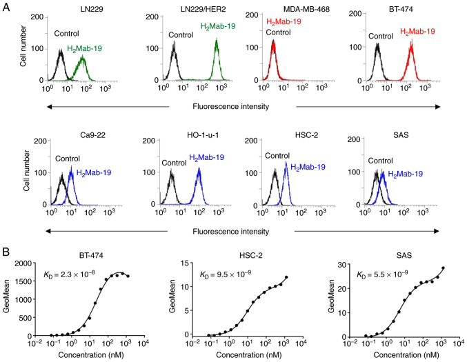 Fig. 4 Characterization of H2Mab-19 using flow cytometry. (A) Glioblastoma cell lines (LN229, LN229/HER2), breast cancer cell lines (MDA-MB-468, BT-474), and oral cancer cell lines (Ca9-22, HO-1-u-1, HSC-2, SAS) were treated with H2Mab-19. (B) Determination of binding affinity of H2Mab-19 for BT-474, HSC-2, and SAS using flow cytometry. (Takei J, et al., 2020)
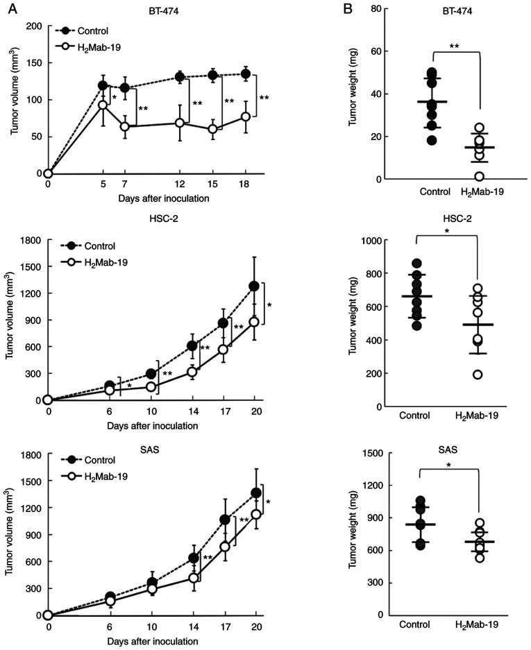 Fig. 5 Evaluation of the antitumor activity of H2Mab-19. (A) Tumor volume and (B) tumor weight were measured from BT-474 (upper), HSC-2 (middle), and SAS (lower) xenografts. (Takei J, et al., 2020)
The flow cytometer measures total fluorescence signals from each cell, while the image-based system acquires images and analyzes fluorescently stained autophagosomes within the cells, which can provide more accurate measurements of autophagy activity.
Ask a Question
Average Rating: 4.0 | 1 Scientist has reviewed this product
Convenient
The tumor cell product's convenient shipping options and efficient customer service have made our procurement process smooth and hassle-free.
19 Apr 2023
Ease of use
After sales services
Value for money
Write your own review
- You May Also Need

