Featured Products
Our Promise to You
Guaranteed product quality, expert customer support

ONLINE INQUIRY
SUSA
Cat.No.: CSC-C6210X
Species: Human
Source: testicular germ cell tumor
Morphology: epitheloid cells growing adherently in monolayers
Culture Properties: monolayer
- Specification
- Background
- Scientific Data
- Q & A
- Customer Review
Immunology: cytokeratin +, cytokeratin-7 +, cytokeratin-8 +, cytokeratin-17 -, cytokeratin-18 +, cytokeratin-19 +, desmin -, endothel -, EpCAM +, GFAP -, neurofilament -, vimentin +
Viruses: PCR: EBV -, HBV -, HCV -, HIV
The SUSA cell line originates from the malignant testicular teratoma of a 30-year-old male patient which was moderately malignant type B with extensive necrosis and was established in 1977. Within the male reproductive system, the testis functions as an essential organ that produces sperm and secretes androgens. Testicular teratoma originates from germ cells and usually presents multiple tissue types including epithelium, glands, neural tissue and muscle. The SUSA cells demonstrate epithelial morphology and form monolayer cultures while showing typical epithelial cell behaviors when grown in vitro.
Researchers use this cell line extensively to examine the molecular mechanisms that drive testicular cancer through gene expression analysis, regulatory processes in signaling pathways, and drug resistance studies. Research shows that the SUSA cell line demonstrates increased IGF1R signaling pathway expression and is crucial for platinum drug resistance. Furthermore, the cell line functions as a model system to evaluate the therapeutic effectiveness and action mechanisms of chemotherapeutic agents while specifically investigating cisplatin and etoposide cross-resistance.
Apoptosis in Testis and Bladder Cancer Cells Following Cisplatin Treatment
Metastatic testicular germ cell tumors (TGCT) show an over 80% cure rate with cisplatin-based therapy, unlike most solid metastatic cancers resistant to chemotherapy. TGCTs are hypothesized to be sensitive due to reduced DNA repair capability and a higher tendency to undergo apoptosis, particularly involving non-mutated p53. Köberle et al. explored the role of apoptotic pathways in this sensitivity by examining cisplatin-induced activation in TGCT cells compared to resistant bladder cancer cells. Cisplatin-induced apoptosis was studied in three testis tumor cell (TTC) lines (8333, SuSa, GCT27) and three bladder cancer cell lines (MGH-U1, HT1376, RT112). After 1-hour exposure to cisplatin, the sub-G1 fraction indicating apoptosis was measured. TTC lines showed a stronger dose- and time-dependent apoptosis compared to bladder cancer cells. At 10 and 20 µM, testis tumor cells had significantly more apoptotic cells than bladder cancer cells (P = 0.0219) (Fig. 1A). From 48 hours post-treatment, apoptosis was significantly higher in testis cells with P-values of P = 0.0376 (48 h), P = 0.0063 (72 h), P = 0.0014 (96 h), and P = 0.0082 (120 h) (Fig. 1B). Greater PARP-1 cleavage further indicated higher apoptosis in TTC (Fig. 1C). These results align with previous findings of cisplatin hypersensitivity in TTC due to persistent DNA damage from reduced ICL repair (Fig. 1C).
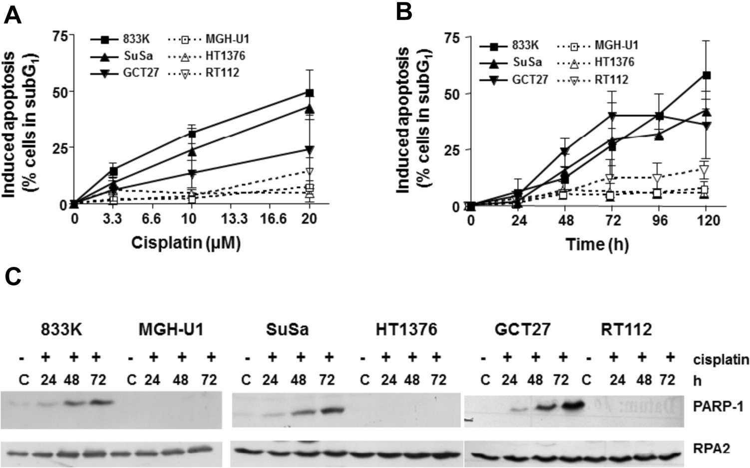 Fig. 1. Determination of apoptosis in testis (filled symbols) and bladder (open symbols) cancer cells after treatment with cisplatin (Köberle B, Usanova S, et al., 2024).
Cell Killing Efficacy of Brentuximab Vedotin in Correlation with CD30 Expression
Testicular germ cell tumors (TGCTs), the most common cancer in young men, are generally treatable with chemotherapy. However, cisplatin-resistant TGCTs in young patients remain a challenge, prompting the need for new therapeutic approaches. Brentuximab vedotin, which targets CD30-a protein overexpressed in many TGCTs-offers a promising treatment option. Yeste-Velasco et al. evaluated the efficacy of brentuximab vedotin alone and with other chemotherapy drugs, considering its potential to target CD30, to enhance treatment outcomes for TGCTs. Dose-response curves for brentuximab vedotin were generated for all 12 TGCT cell lines (833K, 833KR, Susa, SusaR, GCT27, GCT27R, GCT44,TERA-1,NTERA-2,NCG2102, 577MF and TCam-2) using MTS assay (Fig. 2A and B). Sensitivity generally correlated with CD30 expression. Highly CD30-expressive 833K cells were the most sensitive (>500 times). However, NCG2012 cells, despite significant CD30 expression, didn’t respond until concentrations exceeded 5000 ng/ml, likely due to non-specific toxicity. TCam-2, with low CD30 expression, also showed no response until over 5000 ng/ml. CD30-negative lines 577MF and GCT44, and low-expressing TERA1, started responding above 1000 ng/ml. At 125,000 ng/ml, nearly all cells were killed in three days, but about 20% of GCT27 and TCAM2 survived.
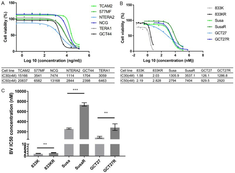 Fig. 2. Brentuximab vedotin dose-response curves of TGCT cell lines (Yeste-Velasco M, Guo T, et al., 2019).
Quality control measures minimize experimental variability, reduce the risk of contamination, and ensure that cell-based assays produce reliable and repeatable results.
Ask a Question
Average Rating: 4.0 | 1 Scientist has reviewed this product
High quality
This product has allowed us to study the behavior of cancer cells in a controlled environment, leading to new insights.
08 Mar 2023
Ease of use
After sales services
Value for money
Write your own review
- You May Also Need
Description: Established from the primary lesion resected from a 76-year-old woman with bulky, moderately differentiated squamous cell carcinoma of the vulva in 1984; cells were reported to express an elevated number of high affinity EGF receptors, to carry gene amplification at 11q13 and to be tumorigenic in nude mice
Description: Established from the solid tumor of a 52-year-old Caucasian man with advanced stage malignant bile duct carcinoma (patient had not received any chemotherapy prior to cell explantation) in 1984 and three consecutive passages in female nu/nu mice (NMRI); primary tumor histology: large cell adenocarcinoma of low differentiation, metastasizing (ascites in patient); human character of cell line confirmed by karyotype in passage #34 of in vitro culture (1986)

Description: Subclone of ACH1P which was established by fusion of the JEG-3-derived HGPRT-defective mutant cell line AC1-1 with primary male trophoblast cells isolated from term human chorion laeve; described as possessing both, a distinct karyotype and a distinct expression pattern of tumor-related genes

Description: One of five phenotypically and cytogenetically distinct subclones of a hybrid (ACH1P) formed by fusing an HGPRT-defective mutant (AC1-1) of JEG-3 human choriocarcinoma with primary human trophoblast (chorion laeve) cells
Description: Established by somatic hybridization of primary male trophoblast cells and the HGPRT-defective choriocarcinoma cell line AC1-1 which was originally derived from cell line JEG-3

Description: NTERA-2 was cloned from cell line TERA-2 which was derived from a metastatic teratocarcinoma of a 22-year-old Caucasian male; cell line also known as NT-2
