- You are here: Home
- Services
- Disease Models
- Orthopedic Disease Models
- Osteoarthritis (OA) Model
- Anterior Cruciate Ligament Transection (ACLT)-Induced Osteoarthritis (OA) Model
Services
-
Cell Services
- Cell Line Authentication
- Cell Surface Marker Validation Service
-
Cell Line Testing and Assays
- Toxicology Assay
- Drug-Resistant Cell Models
- Cell Viability Assays
- Cell Proliferation Assays
- Cell Migration Assays
- Soft Agar Colony Formation Assay Service
- SRB Assay
- Cell Apoptosis Assays
- Cell Cycle Assays
- Cell Angiogenesis Assays
- DNA/RNA Extraction
- Custom Cell & Tissue Lysate Service
- Cellular Phosphorylation Assays
- Stability Testing
- Sterility Testing
- Endotoxin Detection and Removal
- Phagocytosis Assays
- Cell-Based Screening and Profiling Services
- 3D-Based Services
- Custom Cell Services
- Cell-based LNP Evaluation
-
Stem Cell Research
- iPSC Generation
- iPSC Characterization
-
iPSC Differentiation
- Neural Stem Cells Differentiation Service from iPSC
- Astrocyte Differentiation Service from iPSC
- Retinal Pigment Epithelium (RPE) Differentiation Service from iPSC
- Cardiomyocyte Differentiation Service from iPSC
- T Cell, NK Cell Differentiation Service from iPSC
- Hepatocyte Differentiation Service from iPSC
- Beta Cell Differentiation Service from iPSC
- Brain Organoid Differentiation Service from iPSC
- Cardiac Organoid Differentiation Service from iPSC
- Kidney Organoid Differentiation Service from iPSC
- GABAnergic Neuron Differentiation Service from iPSC
- Undifferentiated iPSC Detection
- iPSC Gene Editing
- iPSC Expanding Service
- MSC Services
- Stem Cell Assay Development and Screening
- Cell Immortalization
-
ISH/FISH Services
- In Situ Hybridization (ISH) & RNAscope Service
- Fluorescent In Situ Hybridization
- FISH Probe Design, Synthesis and Testing Service
-
FISH Applications
- Multicolor FISH (M-FISH) Analysis
- Chromosome Analysis of ES and iPS Cells
- RNA FISH in Plant Service
- Mouse Model and PDX Analysis (FISH)
- Cell Transplantation Analysis (FISH)
- In Situ Detection of CAR-T Cells & Oncolytic Viruses
- CAR-T/CAR-NK Target Assessment Service (ISH)
- ImmunoFISH Analysis (FISH+IHC)
- Splice Variant Analysis (FISH)
- Telomere Length Analysis (Q-FISH)
- Telomere Length Analysis (qPCR assay)
- FISH Analysis of Microorganisms
- Neoplasms FISH Analysis
- CARD-FISH for Environmental Microorganisms (FISH)
- FISH Quality Control Services
- QuantiGene Plex Assay
- Circulating Tumor Cell (CTC) FISH
- mtRNA Analysis (FISH)
- In Situ Detection of Chemokines/Cytokines
- In Situ Detection of Virus
- Transgene Mapping (FISH)
- Transgene Mapping (Locus Amplification & Sequencing)
- Stable Cell Line Genetic Stability Testing
- Genetic Stability Testing (Locus Amplification & Sequencing + ddPCR)
- Clonality Analysis Service (FISH)
- Karyotyping (G-banded) Service
- Animal Chromosome Analysis (G-banded) Service
- I-FISH Service
- AAV Biodistribution Analysis (RNA ISH)
- Molecular Karyotyping (aCGH)
- Droplet Digital PCR (ddPCR) Service
- Digital ISH Image Quantification and Statistical Analysis
- SCE (Sister Chromatid Exchange) Analysis
- Biosample Services
- Histology Services
- Exosome Research Services
- In Vitro DMPK Services
-
In Vivo DMPK Services
- Pharmacokinetic and Toxicokinetic
- PK/PD Biomarker Analysis
- Bioavailability and Bioequivalence
- Bioanalytical Package
- Metabolite Profiling and Identification
- In Vivo Toxicity Study
- Mass Balance, Excretion and Expired Air Collection
- Administration Routes and Biofluid Sampling
- Quantitative Tissue Distribution
- Target Tissue Exposure
- In Vivo Blood-Brain-Barrier Assay
- Drug Toxicity Services
Anterior Cruciate Ligament Transection (ACLT)-Induced Osteoarthritis (OA) Model
Looking for a reliable partner to help you evaluate the efficacy of your drug candidates on osteoarthritis (OA)? Look no further than Creative Bioarray. With our years of expertise in OA research, we have established a stable OA model induced by anterior cruciate ligament transection (ACLT) that ensures accurate and reliable results. Trust us to be your CRO partner for your OA drug development needs.
The ACLT model is the earliest well-known model and is currently the most widely used surgical model in OA research. The rationale for using this model is that ACL injury leads to joint destabilization, which in turn results in post-traumatic OA (PTOA). This model mimics the degradation of articular cartilage following ACL rupture. Compared to meniscectomy, the OA lesions in the ACLT model develop more slowly, making this model easier to use in pharmaceutical studies.
Our Anterior Cruciate Ligament Transection (ACLT)-Induced Osteoarthritis (OA) Model
- Available Animal
Rat - Modeling Method
OA is surgically induced in the knee joint. After anesthetization, the surgical area is shaved and disinfected, and the right knee joint is subsequently exposed by dissecting the skin on the inside of the patella along the patellar tendon. The patella is dislocated laterally, the knee is placed in full flexion, and the anterior cruciate ligament is subsequently transected. After that, a positive anterior drawer test is performed to ensure complete transection of the ligament. - Endpoints
- Body weight
- Behavioral tests
- Histology analysis (knee joint): H&E staining, Toluidine blue staining, Safranin O/Fast green staining
- Cytokine analysis
- Other customized endpoints
Example Data
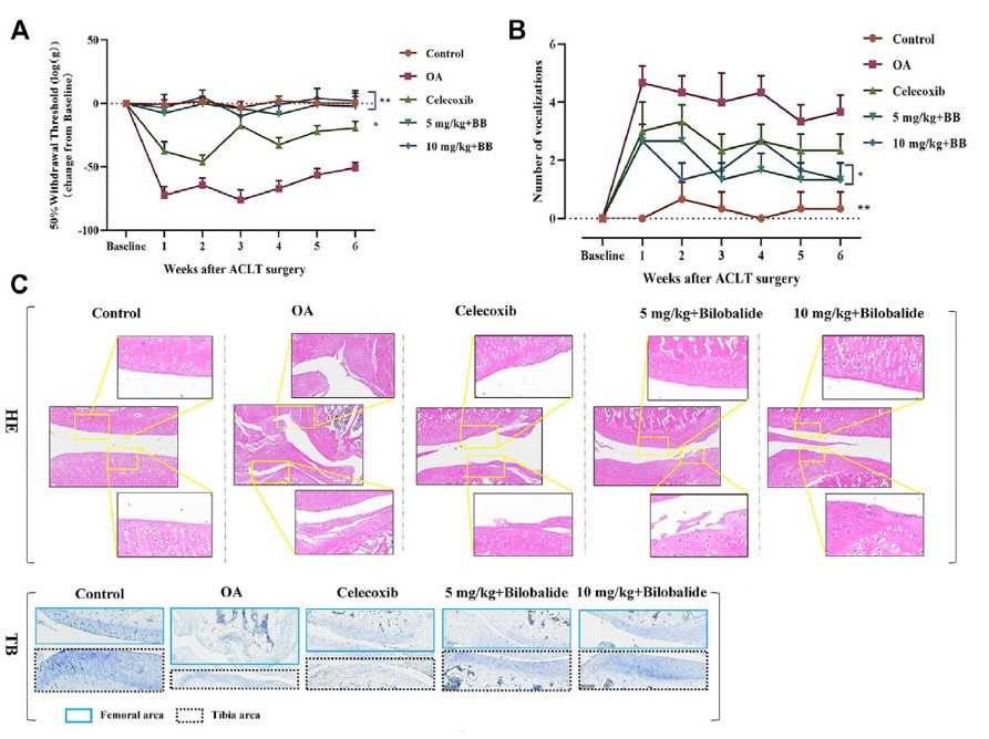
 Fig. 1 Bilobalide attenuates ACLT-induced PTOA in rats via AMPK/SIRT1/mTOR pathway. (A) Mechanical sensitivity. (B) Vocalizations evoked by extension of the knee. (C) Representative images of the knee joint stained with hematoxylin-eosin (HE) and toluidine blue (TB) after 6 weeks of treatment.
Moreover, we also provide other OA models that maybe you are interested in:
- Monosodium Iodoacetate (MIA)-Induced Osteoarthritis (OA) Model
- Papain-Induced Osteoarthritis (OA) Model
- Medial Meniscal Tear (MMT)-Induced Osteoarthritis (OA) Model
Quotation and Ordering
Creative Bioarray has extensive experience and expertise in designing, validating, and analyzing animal efficacy studies. As a reliable partner, we are committed to providing high-quality services to our clients. If you are interested in our services, please feel free to contact us at any time or submit an inquiry to us directly. We look forward to cooperating with you.
References
- Ma, T., et al. Bilobalide exerts anti-inflammatory effects on chondrocytes through the AMPK/SIRT1/mTOR pathway to attenuate ACLT-induced post-traumatic osteoarthritis in rats. Frontiers in pharmacology, 2022, 13: 783506.
- Kubomura, D., et al. Evaluation of the chondroprotective action of N-acetylglucosamine in a rat experimental osteoarthritis model. Experimental and Therapeutic Medicine, 2017, 14(4): 3137-3144.
- Kuyinu, E.L., et al. Animal models of osteoarthritis: classification, update, and measurement of outcomes. Journal of orthopaedic surgery and research, 2016, 11: 1-27.
Explore Other Options
For research use only. Not for any other purpose.

