Featured Products
Our Promise to You
Guaranteed product quality, expert customer support

ONLINE INQUIRY

- Specification
- Background
- Scientific Data
- Q & A
- Customer Review
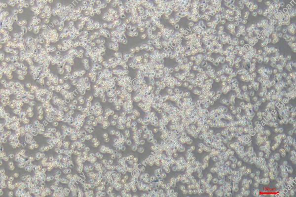
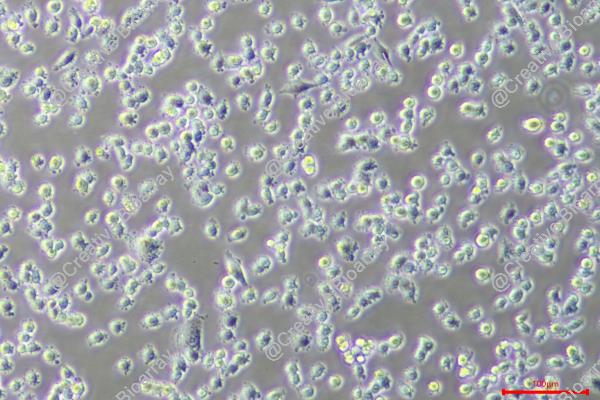
IHH-4 cell line development stems from a lymph node metastasis of papillary thyroid carcinoma found in a 75-year-old male patient. Papillary thyroid carcinoma represents the predominant thyroid cancer type with generally positive outcomes but lymph node metastases can occur in some patients. Sekiguchi, M., together with his team created this cell line which researchers commonly used for thyroid cancer studies. IHH-4 cells demonstrate epithelial morphology while maintaining strong surface adhesion properties and expressing typical cancer cell features. Researchers use these cells to explore how thyroid cancer metastasizes and to understand its biological behavior through cell growth patterns, programmed cell death, and molecular signaling processes. For example, studies indicated that reducing LINC00152 expression blocks both cell growth and movement in IHH-4 cells while increased LINC00152 expression promotes these activities. Additionally, the TGF-β1 treatment causes significant downregulation of lncRNA-NEF which results in decreased migration and invasion abilities of IHH-4 cells. Researchers also use the IHH-4 cell line to study how different drugs affect thyroid cancer cells. For example, aloperine triggers the death of IHH-4 cells by activating the caspase pathway.
Neferine Inhibits the Growth of Thyroid Cancer Cells
Thyroid cancer is rising in prevalence and presents treatment challenges, especially its undifferentiated forms. Neferine possesses various pharmacological activities, which have been applied in diverse disease models, including various tumors. To assess neferine's effect on thyroid cancer, IHH-4 and CAL-62 cells were treated with varying neferine concentrations (0-80 μM) for 96 hours (Fig. 1a and b). CCK-8 results revealed dose-dependent inhibition of cell viability, particularly between 5-80 μM (Fig. 1c). The IC50 values for IHH-4 and CAL-62 were 9.47 and 8.72 μM, respectively. Thus, 5 and 10 μM concentrations were used for further tests. Both concentrations significantly reduced cell viability at 96 hours (Fig. 1d). Additionally, they increased apoptosis rates and altered protein expressions: decreased Bcl-2, increased Bax and cleaved caspase-3 (Fig. 1e-i). Neferine also lowered Ki67 expression levels. Overall, neferine inhibited growth and promoted apoptosis in thyroid cancer cells.
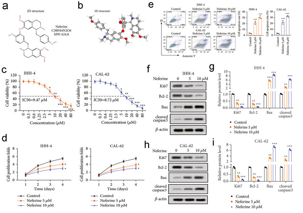 Fig. 1. Neferine suppressed the proliferation and enhanced apoptosis of thyroid cancer cells (Li S, Zhang Y, et al., 2022).
Forced miR-671-5p Repressed Progression of Papillary Thyroid Carcinoma
Papillary thyroid carcinoma (PTC) shows a rise in occurrence and metastasis potential but lacks early diagnostic markers. Potential therapeutic targets for cancer treatment include dysregulated miRNAs such as tumor-suppressive miR-671-5p which has connections to multiple cancers and oncogenic TRIM14 which functions through STAT3 in papillary thyroid carcinoma. Wang's team transfected mimic of miR-671-5p into IHH-4 and TPC-1 cells to analyze its effects on papillary thyroid carcinoma progression. The qRT-PCR analysis showed that miR-671-5p expression levels increased following transfection with the mimic compared to mock or NC mimic transfections (Fig. 2A). The increased expression of miR-671-5p led to reduced viability of both IHH-4 and TPC-1 cells (Fig. 2B). The miR-671-5p overexpression reduced the cell colony formation ability as shown in Figure 2C. These findings show that miR-671-5p can play a role in inhibiting the growth of papillary thyroid carcinoma cells. Beyond its antiproliferative effects miR-671-5p demonstrated suppression of cell migration (Fig. 3A) and invasion (Fig. 3B). The anti-invasive effect of miR-671-5p on papillary thyroid carcinoma became evident through experimental analysis of IHH-4 and TPC-1 cells. The inhibition of miR-671-5p led to decreased proliferation along with reduced migration and invasion of papillary thyroid carcinoma cells.
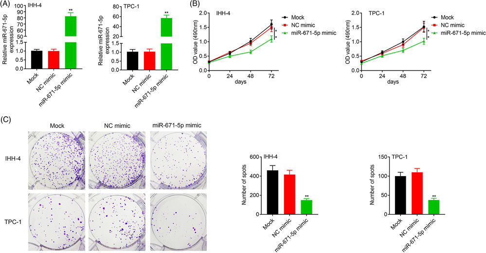 Fig. 2. Transfection with miR-671-5p mimic in IHH-4 and TPC-1 increased miR-671-5p expression (A) and decreased cell viability and proliferation (B-C) compared with that of NC mimic (Wang W, Yuan Y, et al., 2021).
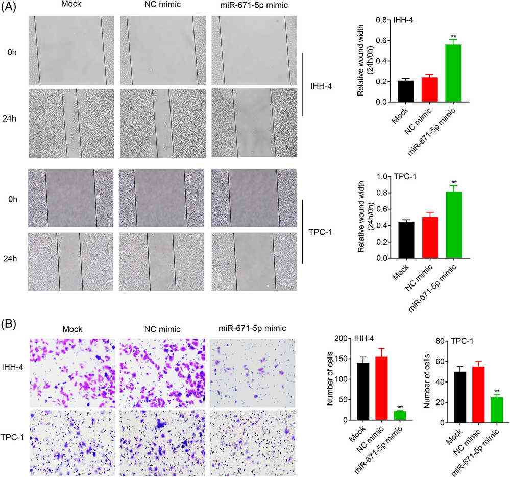 Fig. 3. Transfection with miR-671-5p mimic in IHH-4 and TPC-1 decreased cell migration (A) and invasion (B) compared with that of NC mimic (Wang W, Yuan Y, et al., 2021).
Primary tumor cells refer to the cells forming the initial tumor at the site of origin, while metastatic tumor cells are cells that have spread from the primary tumor to other parts of the body. Metastatic tumor cells often exhibit different characteristics and may be more resistant to treatment than primary tumor cells.
Ask a Question
Average Rating: 5.0 | 1 Scientist has reviewed this product
A trusted supplier
Creative Bioarray is a trusted supplier for obtaining cells with consistent quality.
11 Aug 2023
Ease of use
After sales services
Value for money
Write your own review
- You May Also Need
Description: FTC-133 was isolated from a lymph node metastasis of a follicular thyroid carcinoma. The morphology differs from flat polygonal to spindle formed cells. They retained differentiated thyrocyte function and thyrocyte responsiveness to thyrotropin and local active growth factors. Thyroglobulin immunoreactivity could be shown in the cytoplasm and EGF receptor immunoreactivity could be visualised on the cell membranes. Complex chromosomal changes were detected as well as a p53 mutation.

Description: Species: human male 42 years old;
Tissue: thyroid;
Tumor: carcinoma, follicular;
Derived from: lung metastasis


Description: Established from the primary tumor of a 67-year-old woman with primary thyroid carcinoma (undifferentiated carcinoma, composed of spindle, polygonal and giant cells)
Description: Established from the sarcoma of the thyroid gland from a 47-year-old woman in 1985

Description: Established from the tumor tissue of a 76-year-old woman with metastasizing papillary thyroid carcinoma in 1992
