Featured Products
Our Promise to You
Guaranteed product quality, expert customer support

ONLINE INQUIRY
- Specification
- Background
- Scientific Data
- Q & A
- Customer Review
MA-104 cells were initially isolated from embryonic kidneys of African green monkeys using tissue culture, and spontaneously immortalised to produce a stable and easy-to-cultivate cell line.MA-104 cells are epithelial and adherent growth-like cells. They can double within 2–3 days, and grow fairly quickly, which makes them an excellent candidate for a large scale lab culture.
More specifically, the MA-104 cell line is highly susceptible to a variety of viruses, including monkey rotavirus (Simian rotavirus SA11) and African swine fever virus (ASFV). This attribute makes it useful for viral transmission, viral disease research and vaccine development. When rotavirus infected MA-104 cells, for instance, the virus replicated and spread rapidly in the cells, resulting in clear cytopathic phenomena (CPE) including crumpling, rounding and shedding. By observing these CPE features, researchers can perform qualitative and quantitative studies of rotaviruses. This includes virulence determination of the virus, evaluation of immunogenicity during vaccine development, and screening experiments for antiviral drugs. In addition, due to its cross-species adaptability and high viral load, the MA-104 cell line is widely used in many research institutions around the world and is an indispensable biological tool for virology research.
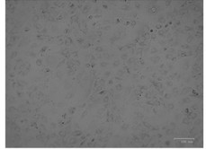 Fig. 1. MA-104 cells (Rai A, Pruitt S, et al., 2020).
ALO Inhibited ASFV Replication In Vitro
ASF is a double-stranded DNA virus transmitted through arthropod vectors. African swine fever (ASF) has become a global pandemic due to inadequate prevention and control measures, posing a significant threat to the swine industry. The virus's mammalian makeup makes it challenging to create vaccines, and currently no antiviral drug against ASF virus (ASFV) is in the pipeline.
Aloperine (ALO), a quinolizidine alkaloid extracted from the seeds and leaves of bitter beans, exhibits various biological functions, including anti-inflammatory, anti-cancer, and antiviral activities. In this study, Geng's team verified the anti-ASFV activity of ALO in vitro and explored the underlying mechanisms of its action. First, the anti-ASFV activity of ALO (Fig. 1A) was evaluated in MA-104 cells. At 100 μM, ALO showed no cytotoxicity (Fig, 1B). Treatment with varying doses of ALO significantly decreased the fluorescence intensity of ASFVGZ-infected cells compared to DMSO (Fig. 1C). Western blot and qPCR showed dose-dependent reductions in ASFV proteins p72 and p30, and in viral gene copies, with 20 μM ALO reducing A137R gene copies by 1.24 log10 (p < 0.001) (Fig. 1D, 1E). ALO inhibited ASFV for at least 72 hours post-infection (Fig. 1F). Similar non-cytotoxicity and anti-ASFV activity were observed in PK-15, 3D4/21, and WSL porcine cells (Fig. 1G–L), although WSL cells required a higher ALO concentration for effectiveness. These findings demonstrate ALO's efficacy in inhibiting ASFV replication in vitro.
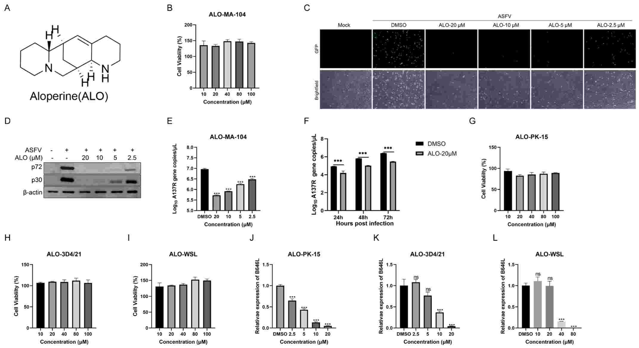 Fig. 1. The cytotoxicity and anti-ASFV activity of ALO were evaluated in African green monkey cells MA-104 (Geng R, Shao H, et al., 2024).
MA-104 as a new substrate for the identification of ASFVs
The African swine fever virus (ASFV), an incredibly deadly virus that is highly infectious among both domestic pigs and wild boars, has been responsible for large-scale outbreaks. The absence of a vaccine has necessitated reliance on quarantine and culling strategies. This diagnosis requires the isolation of ASFV, and the isolation is traditionally made with primary swine macrophages, which are time-consuming and not commercially available. Rai's team suggests that the MA-104 cell line is an inexpensive, reproducible and effective alternative to primary swine macrophages to isolate ASFV from field specimens to the same sensitivity as primary swine macrophages.
Hemadsorption (HA) is the ability of ASFV-infected cells to form rosettes in the presence of swine red blood cells. Once infected with MA-104 cells, rosettes appeared similar to those observed in primary swine macrophages following treatment with ASFV-G or BA71 (Fig. 2A). HA demands a functional CD2-like gene, and isolated CD2-mutated strains fail to form rosettes. However, MA-104 cells lacked non-HA strains and showed clearly positive staining for ASFV protein p30 using immunocytochemistry (Fig. 2B). In swine macrophages, a low background made conclusions ambiguous. In terms of sensitivity to infectious ASFV detection, MA-104 cells gave virus titers around 1 log10 less than swine macrophages, suggesting they could serve as secondary substrates (Fig. 3A). This means that MA-104 cells could also detect ASFV. This would simplify the diagnosis and management of ASFV, offering a key weapon in the fight against the disease.
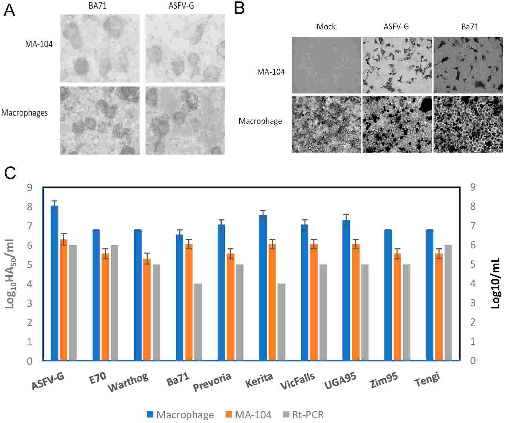 Fig. 2. (A) Hemadsorption was observed 24 h after infection. (B) The presence of the virus was determined by using a monoclonal antibody that detects ASFV protein p30. (C) Comparison of the titration of African Swine Fever Virus (ASFV) isolates in swine macrophages (blue) or MA-104 (orange) cells with real-time PCR (Rai A, Pruitt S, et al., 2020).
Ask a Question
Write your own review
- You May Also Need

