Featured Products
Our Promise to You
Guaranteed product quality, expert customer support

ONLINE INQUIRY
TGBC11TKB
Cat.No.: CSC-C6413J
Species: Human
Morphology: epithelial-like
Culture Properties: Adherent cells
- Specification
- Background
- Scientific Data
- Q & A
- Customer Review
Store in liquid nitrogen.
TGBC11TKB cells were established from lymph node metastasis in a patient suffering from gastric cancer. Lymph nodes are often the first sites of metastasis in gastric cancer due to the cancer's tendency to disseminate through the lymphatic system. The isolation and subsequent culture of cells from these metastatic lymph nodes allow for the creation of a stable cell line that mirrors the genetic and phenotypic attributes of the originating tumor.
The establishment of the TGBC11TKB cell line marks a significant milestone in gastric cancer research. By offering a reliable and well-characterized model system, TGBC11TKB cells enable scientists to dissect the complex molecular and cellular mechanisms underpinning gastric cancer metastasis. This facilitates the development of more effective diagnostic tools and therapeutic strategies.
Analysis of SET7/9 Mutation in TGBC11TKB Cells
SET7/9, a histone methyltransferase, has two distinct functions for lysine methylation. To clarify whether or not SET7/9 is related to carcinogenesis, alterations of SET7/9 in gastric cancers (GCs) were studied. Genetic alterations of SET7/9 were examined in 12 GC cell lines and 25 primary GC tissues. By long RT-PCR from exons 1-8 and PCR-SSCP, TGBC11TKB cells exhibited a deletion of exon 3 and a C to T transition at the first nucleotide of exon 3 in SET7/9 mRNA and genomic DNA, respectively (Fig. 1), which predicts the encoding of a truncated SET7/9 protein. Except for TGBC11TKB cells, no mutation was detected in any cases examined.
 Fig. 1 Longer RT-PCR of SET7/9 exons 1-8 in TGBC11TKB and KATO-III cells (left). Sequencing analysis of the PCR product of SET7/9 in TGBC11TKB cells. After the subcloning of the RT-PCR product showing an abnormal SET7/9 transcript in TGBC11TKB cells, sequencing was performed (right). Deletion of exon 3 predicts encoding a truncated SET7/9 protein lacking the SET domain. (Keller S, et al., 2017)
TP53TG1 Shows Cancer-Specific DNA Methylation-Associated Transcriptional Silencing
The DNA methylation-associated silencing of TP53TG, a lncRNA, produces aggressive tumors that are resistant to cellular death when DNA-damaging agents and small targeted molecules are used. To further demonstrate the silencing of TP53TG1 in cancer cells in association with the presence of promoter CpG island hypermethylation, its transcription start site (TSS) and DNA methylation patterns proceeded to characterize. Bisulfite genomic sequencing of multiple clones confirmed the dense methylation of the CpG island in HCT-116 cells and its unmethylated status in normal colorectal mucosa (Fig. 2A). DNA methylation analyses were extended to another 12 gastrointestinal cancer cell lines (Fig. 2A). In addition to HCT-116, TP53TG1 5'-end CpG island hypermethylation was also found in the colorectal cancer cell line KM12 and the gastric cancer cell lines KATO-III and TGBC11TKB. All of the other cell lines were unmethylated at this locus (Fig. 2A). Importantly, the use of the demethylating agent 5-aza-2'-deoxycytidine restored TP53TG1 expression in the hypermethylated HCT-116, KM12, KATO-III, and TGBC11TKB cell lines (Fig. 2B). Subcellular fractioning showed that the TP53TG1 transcript in DKO cells, in addition to being present in both the cytoplasm and the nucleus, was particularly enriched in the cytosolic compartment (Fig. 2C). RNA fluorescence in situ hybridization (RNA-FISH) corroborated this intracellular localization (Fig. 2C).
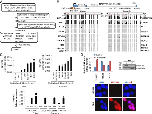 Fig. 2 Epigenetic silencing of the lncRNA TP53TG1 in cancer cells. (A) Bisulfite genomic sequencing analysis of TP53TG1 promoter CpG island in cancer cell lines and normal tissues. (B) DNA methylation-associated transcriptional silencing of TP53TG1 in cancer cells. (C) TP53TG1 RNA-FISH and intracellular localization. (Diaz-Lagares A, et al., 2016)
Common methods include scraping, repeated apposition, enzymatic digestion and others.
Unlike primary tumor samples, the TGBC11TKB cell line provides a renewable, consistent, and well-characterized model system for conducting controlled experiments related to the metastatic process in gastric cancer.
Cell lines derived from metastatic tumors, such as TGBC11TKB, provide a consistent and reproducible model system for studying the complex mechanisms of cancer metastasis. This allows researchers to conduct controlled experiments and make more reliable observations compared to using primary tumor samples or animal models.
The recommended incubation medium for TGBC11TKB cells is DMEM + 5% h.i. FBS.
Ask a Question
Average Rating: 5.0 | 3 Scientist has reviewed this product
Reasonable price
The pricing of the cell products was reasonable and competitive, especially considering the high quality of the cells. I would highly recommend this biological company to other researchers.
17 Mar 2022
Ease of use
After sales services
Value for money
Satisfied
I recently started using the TGBC11TKB cell line from Creative Bioarray for some new gastric cancer studies in my lab, and I've been extremely satisfied with the results.
11 Mar 2024
Ease of use
After sales services
Value for money
Good stability
The cells have retained their phenotypic and genotypic stability, even after repeated passaging.
23 Apr 2024
Ease of use
After sales services
Value for money
Write your own review
- You May Also Need

